
Page 776 - Adams and Stashak's Lameness in Horses, 7th Edition
P. 776
742 Chapter 5
VetBooks.ir
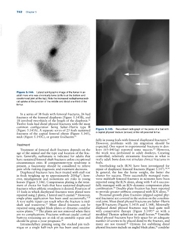
Figure 5.144. Lateral scintigraphic image of the femur in an
adult mare who was chronically lame (stifle is at the bottom and
coxofemoral joint at the top). Note the increased radiopharmaceuti-
cal uptake at the junction of the middle and distal one‐third of the
femur.
In a series of 38 foals with femoral fractures, 26 had
fractures of the femoral diaphysis (Figure 5.143B), and
18 involved two‐thirds of the length of the diaphysis.
36
Twelve foals had distal physeal fractures with the most
common configuration being Salter–Harris type II
(Figure 5.143A). A separate series of 25 foals sustained Figure 5.145. Recumbent radiograph of the pelvis of a foal with
fractures of the capital femoral physis (Figure 5.145), a capital physeal fracture (arrows) of the left proximal femur.
neck (Figure 5.143C), or greater trochanter. 41
fully in young foals with femoral diaphyseal fractures.
85
Treatment However, problems with pin migration should be
expected. One report in experimental fractures in don-
90
Treatment of femoral shaft fractures depends on the keys (65–140 kg) reported some success. However,
age of the animal and the type and location of the frac- the work was performed in adult donkeys. Creating
ture. Generally, euthanasia is indicated for adults that controlled, relatively atraumatic fractures in what is
have sustained femoral shaft fractures unless exceptional really adult bone does not simulate clinical fractures in
circumstances exist. If compartment‐type syndrome is foals.
present, a fasciotomy should be considered to relieve Interlocking nails (ILN) have been investigated for
pain while making diagnostic and treatment decisions. repair of diaphyseal femoral fractures (Figure 5.147). 58,70
Diaphyseal fractures have been treated with stall rest In general, the less the horse weighs, the better the
57
in foals weighing up to approximately 200 kg ; how- chance for success. Three successfully managed trans-
ever, misalignment and nonunions are risks in young verse midshaft femoral fractures in neonates have been
horses (Figure 5.146). Compression plating is the treat- reported using the ILN alone, along with 4 of 6 success-
ment of choice for foals that have sustained diaphyseal fully managed with an ILN–dynamic compression plate
fractures when athletic soundness is desired. Fourteen of combination. Double‐plate fixation has been reported
99
15 foals in which diaphyseal fractures were plated were to provide greater stiffness compared with ILN alone. 70
36
repaired using 2 plates, 1 lateral and 1 cranial. However, Proximal growth plate fractures (slipped capital phy-
single‐plate application has been used occasionally. 7,36 seal fractures) are covered in the section on the coxofem-
A very stable repair can result when the fracture is mid- oral joint. Most distal physeal fractures are Salter–Harris
shaft and transverse. More distal fractures can be type II fractures (Figures 5.143A and 5.148). Minimally
97
repaired using angled blade plates or dynamic condylar displaced distal physeal fractures may heal quite well
screw plates. 11,36 The plates are not removed when there with conservative therapy (Figure 5.149) or with a
35
48
are no complications. Fractures without caudal cortical modified Thomas splint/cast in small horses. Unstable
buttress remaining are at risk of an unstable repair and distal physeal fractures have little space for an adequate
should be given a poor prognosis. 36 number of screws to be placed distal to the fracture, and
Intramedullary pinning using the stacked pin tech- many are not treated. Options for stabilizing distal
36
nique or a single half‐inch pin has been used success- physeal fractures include an angled blade plate, condylar
36