
Page 780 - Adams and Stashak's Lameness in Horses, 7th Edition
P. 780
746 Chapter 5
However, in the acute case, it can be more difficult to Treatment
diagnose due to the amount of swelling and pain that Treatment is often based on chronicity of the lesion.
VetBooks.ir (Figure 5.152), ultrasound, thermography, and local In general, the more acute the lesion, the more the clini-
can occur in the gaskin region. Nuclear scintigraphy
cian can attempt to minimize the formation of a fibrotic
infiltration of the area may all be necessary in horses
with acute injuries. 17 scar (Figure 5.153). Once the mature scar tissue is pre-
sent, then surgery should be considered.
Lateral
A B
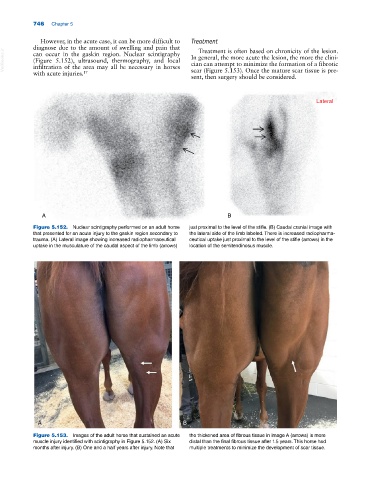
Figure 5.152. Nuclear scintigraphy performed on an adult horse just proximal to the level of the stifle. (B) Caudal cranial image with
that presented for an acute injury to the gaskin region secondary to the lateral side of the limb labeled. There is increased radiopharma-
trauma. (A) Lateral image showing increased radiopharmaceutical ceutical uptake just proximal to the level of the stifle (arrows) in the
uptake in the musculature of the caudal aspect of the limb (arrows) location of the semitendinosus muscle.
A B
Figure 5.153. Images of the adult horse that sustained an acute the thickened area of fibrous tissue in image A (arrows) is more
muscle injury identified with scintigraphy in Figure 5.152. (A) Six distal than the final fibrous tissue after 1.5 years. This horse had
months after injury. (B) One and a half years after injury. Note that multiple treatments to minimize the development of scar tissue.