
Page 782 - Adams and Stashak's Lameness in Horses, 7th Edition
P. 782
748 Chapter 5
FEMORAL NERVE PARALYSIS (CRURAL
PARALYSIS)
VetBooks.ir femoris group of muscles. This muscle group is com-
Paralysis of the femoral nerve affects the quadriceps
posed of the rectus femoris muscle, vastus lateralis mus-
cle, vastus medialis muscle, and vastus intermedius
muscle. This large muscular mass covers the front and
sides of the femur and inserts onto the patella to extend
and fix the stifle.
Etiology
Femoral nerve paralysis may arise from trauma or
unknown causes and may be associated with rhabdomy-
olysis. Injury to the nerve may occur from overstretch-
ing the limb during exertion, kicking, slipping, or while
the horse is tied in a recumbent position. It has also been
reported as a complication of general anesthesia. 24
Hemorrhagic neuritis has been described in 1 horse
that was euthanatized due to femoral paralysis.
24
Unaffected horses had no such lesion at necropsy. The
affected horse also had lesions of rhabdomyolysis, and the
urinary system was affected by myoglobinuria. Postmortem
studies with cadavers revealed that the femoral nerves
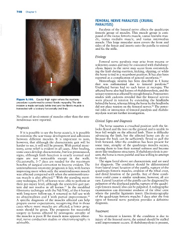
Figure 5.155. Caudal thigh region where the myotomy were placed in tension by extending the hindlimbs
procedure is performed to correct fibrotic myopathy. The skin behind the horse, whereas lifting the horse by the hindlimbs
incision is made vertically (white line) and the fibrotic muscle is did not place tension on the femoral nerves. The poten-
24
transected with a bistoury horizontally (red line). tial roles or interaction of femoral paralysis and rhabdo-
myolysis warrant further investigation.
No cases of involvement of muscles other than the sem-
itendinosus were reported. Clinical Signs and Diagnosis
The horse assumes a crouched position with the fet-
Prognosis locks flexed and the toes on the ground and is unable to
If it is possible to see the horse acutely, it is possible bear full weight on the affected limb. There is difficulty
to minimize the scar tissue development and adhesions advancing the limb, but the affected horse can do so
between different muscles. It is important to note, because the hock can be sufficiently flexed to pull the
however, that although the characteristic gait will be limb forward. After the condition has been present for
harder to see, it will still be present. With partial myec- some time, atrophy of the quadriceps muscles occurs,
tomy, some relief is evident in all cases. After healing, causing them to lose their normal softness and become
some cases develop characteristic, but less pronounced, more like tendinous structures. If rhabdomyolysis is pre-
signs, although limb function is nearly normal and sent, the horse is more painful and less willing to attempt
signs are not noticeable except in the walk. to stand.
Occasionally, 3–7 days are needed for the maximum The signs listed above are characteristic and are used
benefits of surgical correction to become evident. The for diagnosis. The condition should be differentiated
semitendinosus tenotomy prognosis is fair, with horses from lateral (true) luxation of the patella, rupture of the
improving more when only the semitendinosus muscle quadriceps femoris muscles, avulsion of the tibial crest,
was affected compared with when the semimembrano- and distal luxation of the patella. Any of these condi-
sus muscle is also affected. With the standing myot- tions could cause a similar syndrome; however, all are
87
omy technique, 83% of horses were able to perform at rare. Lateral luxation of the patella can be diagnosed by
their pre‐injury level, although the restrictive gait pat- palpation of the displaced patella; rupture of the quadri-
tern did not resolve in all horses. In the modified ceps femoris muscle also can be palpated. A radiographic
52
fibrotomy technique with the Nd:YAG, of the 6 horses examination can determine avulsion of the tibial crest
with long‐term follow‐up, 4 returned to full use with where the patellar ligaments insert. Electromyography
no abnormalities and 1 with partial improvement. of the quadriceps femoris muscles 5 days after the first
44
A specific diagnosis of the muscles affected can help signs of femoral nerve paralysis provides a definitive
prepare owner expectations, recognizing that in those diagnosis.
cases where more muscles are affected, a lower prog-
nosis should be given. The prognosis for successful
surgery in horses affected by neurogenic atrophy of Treatment
the muscles is poor. If the muscle mass appears abnor- No treatment is known. If the condition is due to
mal, nerve conduction studies or electromyography is injury of the femoral nerve, the animal should be stalled
advisable. until improvement occurs. If rhabdomyolysis is present,