
Page 785 - Adams and Stashak's Lameness in Horses, 7th Edition
P. 785
Lameness of the Proximal Limb 751
femoral accessory ligament help to stabilize the cox-
ofemoral joint. 23,45 In addition, the acetabulum is sur-
VetBooks.ir bony margin of the acetabulum.
rounded by a fibrocartilaginous rim that increases the
The accessory ligament is the largest and strongest
45
ligament and is unique to equids. It arises from the fovea
capitis of the femoral head, passes through the acetabular
notch to the pubic groove, and becomes part of the pre-
pubic tendon. The smaller ligament of the head of the
femur (round) arises in the head of the femur adjacent to
the accessory ligament and attaches to the pubic groove.
45
This ligament of the head of the femur can be seen and
debrided arthroscopically, but the accessory ligament is
difficult to visualize. 62–64 The transverse ligament courses
from the outer fibrocartilaginous rim medially across the
45
acetabular notch. The reader is referred to Chapter 1 for
more detailed anatomy of the coxofemoral region.
In general, problems related to the coxofemoral joint
appear to occur most commonly in foals, miniature
horses, and ponies. 23,28,41,53,64 However, horses of any age
may sustain damage to the coxofemoral joint from
trauma. Most conditions are either developmental (oste-
ochondrosis, osteochondritis dissecans, hip dysplasia),
infectious (sepsis involving the coxofemoral joint or
proximal femoral physis), or traumatic (tearing of the
femoral accessory ligament, rupture of the round liga-
ment, capital physeal fractures, hip luxation, IA acetab-
ular fractures, and OA) in origin.
Many of these conditions cause a moderate to severe
lameness and the limb is often outwardly rotated when
viewed from the rear (toe‐out, hock‐in conformation)
(Figures 5.157 and 2.97). In addition, the limb may
appear straighter than the contralateral limb, and the
horse may lean away from the affected limb (Figure 5.142).
External swelling of the coxofemoral joint is often diffi-
cult to detect, but visual enlargement over the greater
trochanter may be evident (Figure 5.158). Pain can often
be elicited with direct inward pressure of this area. Rectal
palpation may reveal swelling along the axial aspect of
the joint but can only be performed in larger horses and
gives many false‐negative results. Crepitus with limb
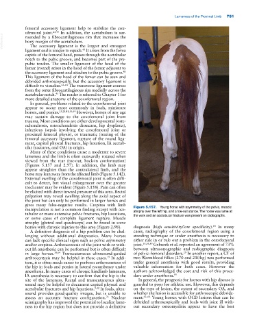
manipulation is not a common finding except with ace- Figure 5.157. Young horse with asymmetry of the pelvis, muscle
atrophy over the left hip, and a toe‐out stance. The horse was lame at
tabular or more extensive pelvic fractures, hip luxations, the walk and an acetabular fracture was present on radiographs.
or some cases of complete ligament rupture. Muscle
atrophy (gluteal and quadriceps) can be found in some
horses with chronic injuries to this area (Figure 2.98). diagnosis (high sensitivity/low specificity). In many
18
A definitive diagnosis of a hip problem can be chal- cases, radiography of the coxofemoral region using a
lenging without additional diagnostics. Many horses standing technique or under anesthesia is necessary to
can lack specific clinical signs such as pelvic asymmetry either rule in or rule out a problem in the coxofemoral
and/or crepitus. Arthrocentesis of the joint with or with- joint. 55,56,89 Geburek et al. reported an agreement of 73%
out IA anesthesia can be performed but is often difficult between ultrasonographic and radiographic diagnosis
29
in large horses. 38,64 Transcutaneous ultrasound‐guided of pelvic–femoral disorders. In another report, a CT of
arthrocentesis may be helpful in these cases. In addi- two Warmblood fillies (270 and 210 kg) was performed
19
tion, it is often much easier to perform arthrocentesis of under general anesthesia with good results, providing
the hip in foals and ponies in lateral recumbency under valuable information for both cases. However the
anesthesia. In many cases of chronic hindlimb lameness, authors acknowledged the cost and risk of this proce-
IA anesthesia is necessary to confirm that the hip is the dure under anesthesia. 95
site of the lameness. Rectal and transcutaneous ultra- In general, the prognosis for horses with hip disease is
sound may be helpful to document capital physeal and guarded to poor for athletic use. However, this depends
acetabular fractures and hip luxations. 9,32 In foals, ultra- on the type of lesion, the extent of secondary OA, and
sound provides good‐quality images, but is unable to whether the lesion is accessible for arthroscopic debride-
assess an accurate fracture configuration. Nuclear ment. 62–64 Young horses with OCD lesions that can be
74
scintigraphy has improved the potential to localize lame- debrided arthroscopically and foals with joint ill with-
ness to the hip region but does not provide a definitive out secondary osteomyelitis appear to have the best