
Page 783 - Adams and Stashak's Lameness in Horses, 7th Edition
P. 783
Lameness of the Proximal Limb 749
intensive care and support is indicated, and repeated accumulation of deposits of calcium phosphate.
86
attempts to use the muscles should be discouraged. Although few have investigated the serum chemistries of
VetBooks.ir Prognosis reported feature of the condition in horses. 43,86
affected horses, hyperphosphatemia has not been a
The prognosis is guarded to unfavorable. In a report
of two cases of post‐anesthetic femoral paralysis, one Clinical Signs and Diagnosis
had concurrent rhabdomyolysis and was euthanatized. Because clinical signs of lameness seldom occur, horses
The other responded to supportive therapy and was may carry the lesion for years before presentation to a hos-
standing well 7 days after the onset. 24,51 The prognosis pital. The masses are generally 3–12 cm in diameter. The
should be withheld until sufficient time has elapsed to skin is freely movable over the density because it lies
determine whether any function will return. beneath the superficial and deep fascia. They are often
attached to the joint capsule of the lateral femorotibial
joint. Diagnosis is confirmed on radiographs (Figure 5.156).
CALCINOSIS CIRCUMSCRIPTA Once dissected free, the lesions are encapsulated in a
Calcinosis circumscripta in the horse is a calcified tough, white, fibrous capsule. 22,43 The interior is com-
mass commonly located on the lateral aspect of the posed of loculations of finely granular, gritty, white,
22
gaskin adjacent to the stifle (Figure 5.156). 22,33,43,86 The paste‐like material. Histologically, the granules are
condition has also been observed in other locations such surrounded by a granulomatous inflammatory reac-
22
as the carpus, tarsus, shoulder, and pectoral region. 22,34,86 tion. Although it is difficult to make a broad compari-
The locations can be unilateral, bilateral, or multiple son, the lesion in humans is believed to be secondary to
locations on the body. 22,43,86 Males appear to be affected collagen necrosis. 91
the most, as only a few of the total cases presented in the
literature are females. 22,33,43,86 The age at presentation
has ranged from 1 to 13 years, with mention that the Treatment
condition has not been reported in foals younger than 6 The only treatment is surgical excision. 22,43 However,
months of age. 22,33,34,43,86 the lesions seldom cause a clinical problem. The loca-
tion is one that tends to dehisce following surgery
43
Etiology and must be taken seriously in that respect. Also, due
to the proximity to the femorotibial joint, there is a
The etiology is unknown. The description of the potential to create a septic arthritis. If surgery is per-
22
lesion best fits that of dystrophic calcinosis character- formed, a tension‐relieving closure using mattress
ized by calcinosis circumscripta. Tumor calcinosis sutures and a sutured stent bandage is advised. A
implies a metabolic derangement in calcium/phosphorus circumferential adherent surgical drape is helpful in pre-
metabolism leading to hyperphosphatemia and resulting venting swelling.
A B
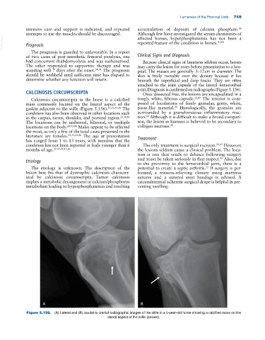
Figure 5.156. (A) Lateral and (B) caudal to cranial radiographic images of the stifle in a 5‐year‐old horse showing a calcified mass on the
lateral aspect of the stifle (arrows).