
Page 781 - Adams and Stashak's Lameness in Horses, 7th Edition
P. 781
Lameness of the Proximal Limb 747
In the acute stages, nonsteroidal anti‐inflammatory reported by Janicek et al. using a Nd:YAG laser to tran-
drugs (NSAIDs) or other pain medication are usually sect fibrotic mass (fibrotomy). Advantages reported
VetBooks.ir gaskin region. It is ideal to try to move and stretch the improved the visualization of the fibrotic mass and facil-
were a more controlled and complete hemostasis, which
required to help minimize the swelling and pain in the
itated the differentiation from normal tissue.
muscles as soon as the horse can tolerate it. The hope is
44
to create as elongated and functional scar of the Bramlage described a semitendinosus tenotomy at
muscle(s) as possible. Initially, the hindlimb can be the level of its insertion on the proximal medial tibia
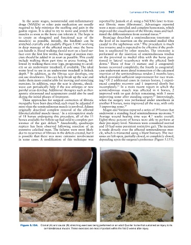
stretched cranially and caudally (Figure 5.154) working caudal to the saphenous vein. The procedure is much
8
in deep massage of the affected muscle once the horse less invasive and is expected to be effective if the prob-
can handle it. Hand walking should start on a hard sur- lem is unaffected by other muscles. The tenotomy is
face over the first few weeks, but range of motion exer- performed at the insertion of semitendinosus tendon
cises should be added in as soon as possible. This could on the proximal to medial tibia with the horse posi-
include walking them part time in arena footing, fol- tioned in lateral recumbency with the affected limb
lowed by walking them over logs, progressing to caval- down. Three of four (1 mature and 2 congenital)
8
etti or an underwater treadmill, if available. The ideal horses recovered completely; the fourth (a congenital)
water level to use in an underwater treadmill is fetlock case underwent more distal transection of the calcaneal
depth. In addition, as the fibrous scar develops, one insertion of the semitendinosus tendon 2 months later,
59
can use shockwave. This can help break up the scar and which provided sufficient improvement for race train-
make them more comfortable for moving and stretching ing. Of 2 additional cases in mature horses, 1 experi-
8
exercises. In addition, once the scar is chronic, shock- enced complete recovery and 1 improved slowly but
wave can periodically help if the size enlarges or new incompletely. In a more recent report in which the
31
painful areas develop. Additional therapies such as ther- semitendinosus muscle was affected in 6 horses, 2
apeutic ultrasound and acupuncture could also be used improved with no gait deficit remaining, with 3 more
during the initial phases of treatment. improving some after standing surgery. Interestingly,
87
Three approaches for surgical correction of fibrotic when the surgery was performed under anesthesia in
myopathy have been described; each must be adjusted if another 8 horses, none improved all the way, with only
more than the semitendinosus muscle is involved. Adams 3 improving some. 87
originally described complete removal of the affected Magee and Vatistas reported a series of 39 horses that
fibrotic/calcified muscle tissue. In a retrospective study underwent a standing focal semitendinosus myotomy.
1
52
of 18 horses undergoing this procedure, all of the 11 Average wound healing time was 4.7 weeks overall.
horses available for follow‐up had mild to complete per- Eighty‐three percent of horses were able to perform at
sistence of the gait deficit. Anecdotally, quadriceps their pre‐injury level. Nineteen were considered normal
96
rupture has been observed following resection of an and 10 had some persistent restrictive gait. The incision
extensive calcified mass. The failures were most likely is made directly over the affected semitendinosus mus-
due to recurrence of fibrosis in the defects created, but it cle, which is transected using a blunt bistoury. The inci-
is possible that there was more extensive involvement sions are left open, partially closed, or completely closed,
in some cases. A modification of this technique was depending upon the surgeon’s preference (Figure 5.155).
A B
Figure 5.154. Cranial (A) and caudal (B) stretching exercises being performed on an adult Quarter horse that sustained an injury to its
semitendinosus muscle. These exercises are most important within the first 2 weeks after injury.