
Page 786 - Adams and Stashak's Lameness in Horses, 7th Edition
P. 786
752 Chapter 5
Diagnosis
Lameness in young horses may be localized to the hip
VetBooks.ir by eliminating the remainder of the limb as the source of
pain along with the characteristic stance of the limb.
Physical examination findings may reveal pain on palpa-
tion and manipulation of the hip. IA anesthetic of the
coxofemoral joint can be performed to aid diagnosis, but
young horses do not tolerate the procedure very well. A
definitive diagnosis is usually achieved via radiography.
Better quality radiographs can generally be obtained
under general anesthesia, although ventrodorsal and
oblique radiographs of the pelvis can be performed in the
standing horse. 55,56,64 Radiographic abnormalities con-
sistent with OCD of the hip are similar to those of other
locations and include SCLs, osteochondral fragments,
abnormal contour of the femoral head or acetabulum,
and shallow and irregular acetabulum (Figure 5.159A).
Treatment
Depending on the severity of the OCD lesion, con-
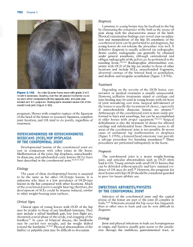
Figure 5.158. An older Quarter horse mare with grade 3 of 5 servative or medical treatment is usually unsuccessful.
hindlimb lameness. Swelling over the left greater trochanter could However, palliative treatments aimed at cartilage and
be seen when compared with the opposite side, and pain was joint healing may be used in young horses with the hope
elicited with firm palpation. Radiographs revealed severe OA of the of joint remodeling over time. Surgical debridement of
coxofemoral joint (Figure 5.163). the lesion is usually the treatment of choice, especially
64
if osteochondritis dissecans lesions are present.
prognosis. Horses with complete rupture of the ligament Arthroscopy of the coxofemoral joint is more easily per-
of the head of the femur or accessory ligament, complete formed in foals and weanlings, but can be accomplished
joint luxation, and OA tend to do poorly, regardless of in older horses with proper equipment. 39,62–64 Surgical
treatment. debridement is also the treatment of choice for affected
cartilage and subchondral bone. However, access to all
areas of the coxofemoral joint is not possible. In severe
OSTEOCHONDROSIS OR OSTEOCHONDRITIS cases of unilateral hip malformation or dysplasia
(Figure 5.159A), a femoral head ostectomy may provide
DISSECANS (OCD)/HIP DYSPLASIA a salvage procedure for breeding soundness. These
80
OF THE COXOFEMORAL JOINT procedures are performed infrequently in the horse.
Developmental lesions of the coxofemoral joint are
rare in comparison with other joints in the horse. Prognosis
Malformation of the joint, hip dysplasia, osteochondri-
tis dissecans, and subchondral cystic lesions (SCLs) have The coxofemoral joint is a major weight‐bearing
been described in the coxofemoral joint. 38,64,65,78,89 joint, and articular abnormalities such as OCD often
lead to OA. Young animals with small OCD lesions that
can be debrided arthroscopically and have minimal evi-
Etiology dence of OA may do well. However, the prognosis for
64
The cause of these developmental lesions is assumed most horses with hip OCD should be considered guarded
to be the same as for other OCD‐type lesions. It is to poor for future athletic use.
unknown why there is a low prevalence of OCD‐type
lesions in the hip compared with other locations. Much
of the coxofemoral joint is weight bearing; therefore, the INFECTIOUS ARTHRITIS/PHYSITIS
development of SCLs could be trauma induced, similar OF THE COXOFEMORAL JOINT
to other weight‐bearing joint surfaces.
Infection of the coxofemoral joint and the capital
physis of the femur are part of the joint ill complex in
Clinical Signs foals. 56,69 Infections around the hip occur less frequently
Clinical signs of young horses with OCD of the hip than at other sites in foals and can be very difficult to
may be similar to those of any hindlimb lameness. They diagnose.
may include a stilted hindlimb gait, low foot flight arc,
shortened cranial phase of the stride, and dragging of the Etiology
hindfeet. In cases of bilateral disease, the hindlimbs
65
may be carried very straight, and the weight shifted Joint and physeal infections in foals are hematogenous
toward the forelimbs. 38,78,89 Physical abnormalities of the in origin, and bacteria usually gain access to the circula-
limb(s) or palpable pain may be difficult to document. tion through the umbilicus, gastrointestinal tract, or