
Page 789 - Adams and Stashak's Lameness in Horses, 7th Edition
P. 789
Lameness of the Proximal Limb 755
and the round ligament both attach to the femoral head
23
and provide the primary stability to the hip. The fibro-
VetBooks.ir lizes the head of the femur within the joint. In horses, the
cartilaginous rim around the acetabulum further stabi-
ilium tends to fracture before the hip luxates. Foals,
38
miniature horses, and ponies are most frequently
affected. 23,38,53,69,72 Coxofemoral subluxation and luxation
are generally unilateral, and the head of the femur nearly
always becomes craniodorsal to the acetabulum. 10,23
Etiology
Both the accessory and the ligament of the head of
38
the femur (round) must rupture for a luxation to occur.
Trauma is nearly always the cause. Violent overexten-
sion and falling on the point of the stifle with the femur
in a vertical position occasionally produce fracture and/
or subluxation or luxation of the coxofemoral joint. 10,38
A tethered horse that catches its foot in a rope or a hal-
ter may dislocate the hip in the struggle to free itself.
Because the acetabulum is deep and the head of the
femur is large, excessive trauma is usually necessary to
dislocate this joint. Fractures of the dorsal rim often
accompany the luxation because of the deep acetabulum
in the horse. Additionally, absence or partial tearing of
38
the ligament of the head of the femur may predispose to
subluxation and luxation with or without associated
trauma. 2,9,38
Coxofemoral luxations may be complicated by
upward fixation of the patella. 16,28,38 The upward patel-
lar fixation may potentially contribute to the luxation
or occur because of it. If it precedes the luxation, the
secondary hip luxation is believed to occur as a result of
the violent contraction of the quadriceps muscles trying
to flex the limb while the stifle is locked in extension.
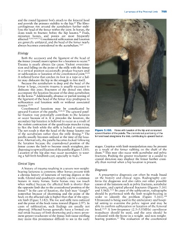
The net result is that the head of the femur luxates out Figure 5.160. Horse with luxation of the hip and concurrent
of the acetabulum rather than the stifle flexing. The upward fixation of the patella. The craniodorsal positioning of the
38
patella usually becomes unfixed at the time of the luxa- femoral head straightens the limb, contributing to upward fixation.
tion. Alternatively, the patella becomes locked following
the luxation because the craniodorsal position of the
femur causes the limb to become much straighter, pre- stages. Crepitus with limb manipulation may be present
disposing to upward fixation of the patella (Figure 5.160). as a result of the femur rubbing on the shaft of the
Luxation of the hip also may occur secondary to wear- ilium. This may also occur with acetabular and pelvic
38
ing a full‐limb hindlimb cast, especially in foals. 94 fractures. Pushing the greater trochanter in a caudal to
cranial direction may displace the femur further crani-
Clinical Signs ally than normal when a hip luxation is present.
A history of trauma resulting in a severe non‐weight‐
bearing lameness is common; other horses present with Diagnosis
a chronic history of lameness of varying degrees at the A presumptive diagnosis can often be made based
walk. Gluteal and quadriceps atrophy can be a common on the history and clinical signs. Radiography con-
feature in many horses. Some horses may toe touch firms the diagnosis and also rules out other possible
when walked because the affected limb is shorter than causes of the lameness such as pelvic fractures, acetabular
the opposite limb due to the craniodorsal position of the fractures, and capital physeal fractures (Figures 5.161
femur. In the case of luxation, the limb may “dangle” and 5.162). 55,56 In cases of the subluxation, radiographs
23
somewhat because of shortening and the point of the should be performed with the limb weight‐bearing in
hock on the affected side is higher than that of the oppo- order to identify the problem (Figure 5.161). 100
site limb (Figure 5.142). The toe and stifle turn outward Ultrasound is being used in the ambulatory and hospi-
and the point of the hock turns inward (Figure 2.97). In tal setting to examine the pelvic region and may be
cases of subluxation, such findings are usually not able to confirm subluxation or luxation of the femoral
observed. Affected horses generally have a limited cra- head. 10,32 A low‐frequency (2.5–5.0 MHz) curvilinear
nial stride because of limb shortening and a more prom- transducer should be used, and the area should be
inent greater trochanter of the femur. Soft tissue swelling evaluated with the horse in a weight‐ and non‐weight‐
may make this prominence difficult to see in the early bearing position. The evaluation of the coxofemoral
10