
Page 793 - Adams and Stashak's Lameness in Horses, 7th Edition
P. 793
Lameness of the Proximal Limb 759
Etiology to diagnose with ultrasound. Both transcutaneous and
transrectal ultrasound should be performed to evaluate
Trauma is the cause in nearly all cases. Stress or fatigue
VetBooks.ir fractures occur elsewhere in the pelvis such as the ilium, not uncommon.
the area. Concurrent fractures of the pelvis and hip are
but are usually not confined to the acetabulum.
23
Treatment
Clinical Signs
Conservative treatment with stall confinement is
Horses with acetabular fractures present with similar often the treatment of choice with acetabular fractures.
clinical signs as horses with pelvic fractures or hip dis- Euthanasia may be indicated with severely displaced
ease. They usually have a significant unilateral lameness fractures because there is no known surgical treatment.
and may have gluteal muscle atrophy if the condition is Small IA fractures from the cranial and caudal perimeter
chronic. Swelling, pain, and crepitus may be palpable of the acetabulum may be removed from the joint with
over the greater trochanter, and there is often pelvic the arthroscope to potentially improve the prognosis.
64
asymmetry if other pelvic fractures are present However, removal of large fragments appears to rarely
(Figure 5.157). A rectal examination may also reveal be successful. Minimally displaced acetabular fractures
swelling axial to the coxofemoral joint, but it may be heal very well with confinement, and horses often can
difficult to detect with acetabulum fractures alone.
achieve their full athletic potential (Figure 5.164).
Diagnosis Prognosis
Nuclear scintigraphy can be valuable in identifying In general, horses with fractures of the acetabulum
that the cause of lameness is in the hip, but ultimately have a worse prognosis for soundness than those with
18
radiography or ultrasound is necessary to document an fractures elsewhere in the pelvis. 23,72,75 However, most
acetabular fracture and rule out other possible problems acetabular fractures respond better to conservative
in the hip and pelvis (Figure 5.164). 23,32,55 Nondisplaced treatment than many other hip problems such as capital
acetabular fractures may be difficult to visualize with physeal fractures, luxation, and round ligament rupture.
ultrasound. However acetabular rim fractures are easier
Approximately 20% of horses with articular fractures
are capable of athletic performance. However, this
76
greatly depends on the fracture configuration and degree
of displacement.
ACKNOWLEDGMENT
The authors thanks Dr. Kenneth E. Sullins and Gary
Baxter for their contributions to this chapter in the pre-
vious edition.
References
1. Adams OR. Fibrotic myopathy in the hindlegs of horses. J Am Vet
Med Assoc 1961;139:1089.
2. Adams OR. Lameness in Horses, 3rd ed. Lea and Febiger,
Philadelphia, 1974;299–303.
3. Ahern BJ, Richardson DW, Boston RC, Schaer TP. Orthopedic
infections in equine long bone fractures and arthrodeses treated
by internal fixation: 192 cases (1990–2006). Vet Surg 2010:39:
588–593.
4. Barcelo Oliver F, Russell TM, Uprichard KL, et al. Treatment of
septic arthritis of the coxofemoral joint in 12 foals. Vet Surg
2017;46:530–538.
5. Bertoni G, Gnudi G, Pezzoli G. Two cases of calcinosis circum-
scripta in the horse. Annali della Facolta di Medicina Veterinaria,
Universita di Parma 1993;13:201–210.
6. Bertoni L, Seignour M, de Mira MC, et al. Fractures of the third
trochanter in horses: 8 cases (2000–2012). J Am Vet Med Assoc
2013;243:261–266.
7. Boulton CH, Dallman MJ. Equine femoral fracture repair: a case
report. J Equine Vet Sci 1983;3:60–64.
8. Bramlage LR, Reed SM, Embertson RM. Semitendinosus tenot-
omy for treatment of fibrotic myopathy in the horse. J Am Vet
Med Assoc 1985;186:565–567.
9. Brenner S, Whitcomb MB. How to diagnose equine coxofemoral
subluxation with dynamic ultrasonography. Proc Am Assoc Equine
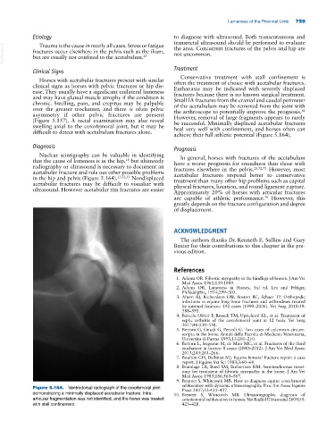
Figure 5.164. Ventrodorsal radiograph of the coxofemoral joint Pract 2007;53:433–437.
demonstrating a minimally displaced acetabular fracture. Intra‐ 10. Brenner S, Whitcomb MB. Ultrasonographic diagnosis of
articular fragmentation was not identified, and the horse was treated coxofemoral subluxation in horses. Vet Radiol Ultrasound 2009;50:
with stall confinement. 423–428.