
Page 790 - Adams and Stashak's Lameness in Horses, 7th Edition
P. 790
756 Chapter 5
VetBooks.ir
A B
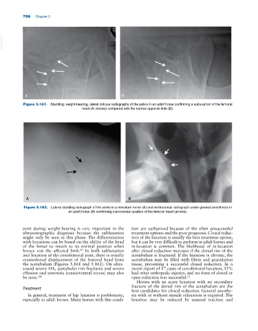
Figure 5.161. Standing, weight‐bearing, lateral oblique radiographs of the pelvis in an adult horse confirming a subluxation of the femoral
head (A; arrows) compared with the normal opposite limb (B).
A B
Figure 5.162. Lateral standing radiograph of the pelvis in a miniature horse (A) and ventrodorsal radiograph under general anesthesia in
an adult horse (B) confirming craniodorsal luxation of the femoral head (arrows).
joint during weight‐bearing is very important in the tion are euthanized because of the often unsuccessful
ultrasonographic diagnosis because the subluxation treatment options and the poor prognosis. Closed reduc-
might only be seen in this phase. The differentiation tion of the luxation is usually the best treatment option,
with luxations can be based on the ability of the head but it can be very difficult to perform in adult horses and
of the femur to return to its normal position when re‐luxation is common. The likelihood of re‐luxation
horses rest the affected limb. In both subluxation after closed reduction increases if the dorsal rim of the
10
and luxation of the coxofemoral joint, there is usually acetabulum is fractured. If the luxation is chronic, the
craniodorsal displacement of the femoral head from acetabulum may be filled with fibrin and granulation
the acetabulum (Figures 5.161 and 5.162). On ultra- tissue, preventing a successful closed reduction. In a
sound severe OA, acetabular rim fractures and severe recent report of 17 cases of coxofemoral luxation, 35%
effusion and synovitis (cranioventral recess) may also had other orthopedic injuries, and no form of closed or
be seen. 100 open reduction was successful. 53
Horses with an acute luxation with no secondary
Treatment fracture of the dorsal rim of the acetabulum are the
best candidates for closed reduction. General anesthe-
In general, treatment of hip luxation is problematic, sia with or without muscle relaxation is required. The
especially in adult horses. Many horses with this condi- luxation may be reduced by manual traction and