
Page 778 - Adams and Stashak's Lameness in Horses, 7th Edition
P. 778
744 Chapter 5
FRACTURES OF THE THIRD TROCHANTER
VetBooks.ir located on the lateral aspect of the proximal third of the
The third trochanter is a prominent tuberosity that is
femur. It provides insertion to the tendon of the gluteus
superficialis muscle, which is mainly a flexor of the cox-
ofemoral joint and abductor of the limb. Due to the
30
superficial location, this tuberosity is prone to trauma
causing fractures or injuries such as avulsion of the ten-
don of the gluteus superficialis muscle. It is an uncom-
6
mon cause of lameness with most horses showing severe
lameness in acute cases. Clinical diagnosis can be
challenging without advanced diagnostic techniques.
Generally, it can be diagnosed with radiographs (50°
cranial, 30° lateral caudomedial oblique, or craniolat-
eral to caudomedial 25° oblique); however in horses
with chronic displaced fractures, scintigraphy and ultra-
sound can help further evaluate the area. 18,76 This type of
fracture is often displaced and heals with a fibrous
union. Treatment consists of conservative management
with rehabilitation including stall rest (1–2 months),
controlled hand walking, and turnout (2–12 months). 6,76
The prognosis for return to athletic activity is good after
an appropriate period of rest and restricted exercise.
6
However, another report showed 8 of 12 horses return-
ing to athletic activity with 2 of these horses returning at
a lower level. 76
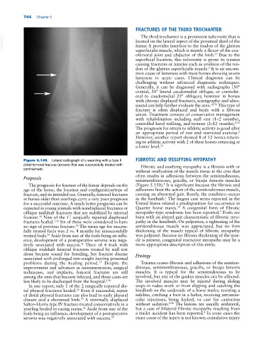
Figure 5.149. Lateral radiograph of a weanling with a type II FIBROTIC AND OSSIFYING MYOPATHY
distal femoral fracture (arrows) that was successfully treated with Fibrotic and ossifying myopathy is a fibrosis with or
confinement.
without ossification of the muscle tissue in the crus that
Prognosis often results in adhesions between the semitendinosus,
semimembranosus, gracilis, or biceps femoris muscles
1
The prognosis for fracture of the femur depends on the (Figure 5.150). It is significant because the fibrosis and
age of the horse, the location and configuration/type of adhesions limit the action of the semitendinosus muscle,
fracture, and its intended use. Generally, femoral fractures causing an abnormal gait. Rarely, the condition occurs
in horses older than yearlings carry a very poor prognosis in the forelimb. The largest case series reported in the
1
for a successful outcome. A much better prognosis can be United States related a predisposition for occurrence in
52
expected in young animals with nondisplaced fractures or Quarter horse mares. A congenital form of fibrotic
8
oblique midshaft fractures that are stabilized by internal myopathy‐type syndrome has been reported. Foals are
fixation. Nine of the 17 surgically repaired diaphyseal born with an altered gait characteristic of fibrotic myo-
36
fractures healed. 7,36 Six of those were considered to have pathy in the hindlimb. On palpation, a tightening of the
no sign of previous fracture. The mean age for success- semitendinosus muscle was appreciated, but no firm
36
fully treated foals was 2 vs. 4 months for unsuccessfully thickening of the muscle typical of fibrotic myopathy
treated foals. Aside from size of the foals being an influ- was palpated. Because no fibrous thickening of the mus-
36
ence, development of a postoperative seroma was nega- cle is present, congenital restrictive myopathy may be a
tively associated with success. Three of 4 foals with more appropriate description of this entity.
36
oblique midshaft femoral fractures treated by stall rest
alone became sound for breeding, but fracture disease Etiology
associated with prolonged non‐weight‐bearing presented
problems during the healing period. Despite the Trauma causes fibrosis and adhesions of the semiten-
57
improvement and advances in instrumentation, surgical dinosus, semimembranosus, gracilis, or biceps femoris
techniques, and implants, femoral fractures are still muscles. It is typical for the semitendinosus to be
among the ones that become infected, and those cases are involved, but any of the gaskin muscles can be affected.
less likely to be discharged from the hospital. 3,50 The involved muscles may be injured during sliding
In one report, only 1 of the 2 surgically repaired dis- stops in rodeo work or from slipping and catching the
tal physeal fractures healed. Even if successful, repair hindlimb on the underside of a horse trailer, resisting a
36
of distal physeal fractures can also lead to early physeal sideline, catching a foot in a halter, receiving intramus-
closure and a shortened limb. A minimally displaced cular injections, being kicked, or cast for castration
50
Salter–Harris type IV fracture treated conservatively in a without sedation. 8,88 The lesions are usually unilateral,
yearling healed to racing status. Aside from size of the but a case of bilateral fibrotic myopathy resulting from
36
foals being an influence, development of a postoperative a trailer accident has been reported. In some cases the
8
seroma was negatively associated with success. 36 exact cause of the injury is not known; cumulative injury