
Page 847 - Adams and Stashak's Lameness in Horses, 7th Edition
P. 847
Principles of Musculoskeletal Disease 813
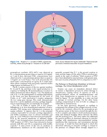
VetBooks.ir IL-1
IL-1
IL-1 receptor
Inducible signaling pathways
Transcriptional and
posttranscriptional
regulation
MMPs PGE 2
Aggrecanase
Figure 7.10. Diagram of IL‐1 activation of MMPs, aggrecanase, brane. Source: Redrawn from figure 6, McIlwraith. Reproduced with
64
and PGE release acting through IL‐1 receptors on the cell mem- permission of American Association of Equine Practitioners.
2
proteoglycan synthesis (42%–64%) was observed at generally accepted that IL‐1 is the pivotal cytokine in
IL‐1 concentrations greater than or equal to 0.01 ng/mL early and late stages of OA, while TNFα is involved pri
at 2 and 4 days. Increased PGE concentrations were marily in the onset of arthritis. Both receptors of TNF
29
2
observed at IL‐1 concentrations greater than or equal to have been identified in synovial tissue, with greater num
1.0 ng/mL at 2 and 4 days. This work showed that the bers seen in joints affected by RA in comparison to OA. 19
much lower concentrations of equine IL‐1 could cause
these effects compared with previously reported studies The Pathways to Morphologic Breakdown of Articular
using human recombinant IL‐1. 104 Cartilage: The Critical Manifestation of OA
The IL‐1 system consists of the two agonist members
IL‐1α and IL‐1β, and these evoke signal production in Trauma can cause an immediate physical defect
response to binding IL‐1R transmembrane receptors to or initiate a degradative process by direct damage to
induce downstream effects (Figure 7.10). The significant chondrocytes, causing release of enzymes as well as
role of IL‐1 in the pathogenesis of cartilage degradation cytokine‐initiated release of metalloproteinases and
in the horse was best demonstrated by the work of PGE from chondrocytes in response to IL‐1. As out
2
Frisbie et al. with interleukin‐1 receptor antagonist lined in Figure 7.8, any instability in a joint can lead
27
(IL‐1ra) using gene therapy. This study demonstrated to damage of normal cartilage. On the other hand,
that if IL‐1 can be inhibited, articular cartilage degrada cartilage compromised by loss of GAG or collagen is
tion in experimental OA can essentially be stopped. vulnerable to normal forces.
The role of TNFα in equine OA is less certain. As has been previously discussed, as cartilage is
Billinghurst et al. first demonstrated induction of intra‐ compressed, the collapse of the collagen network is coun
articular TNF during acute inflammatory responses in tered by the resistance offered by the proteoglycan gel.
equine arthritis. It appears that IL‐1 is the principal The precise nature of the forces exerted through the net
5
cytokine responsible for articular cartilage degradation work is complex and related to both mechanical entrap
and TNFα contributes more to clinical morbidity and ment and chemical bonding effects between the fibers
7
pain. IL‐1 and TNFα have been demonstrated using RT‐ and the proteoglycans. The same author has addressed
PCR in the synovial membrane of inflamed equine the general problem of abnormal softening in articular
joints, and increased serum concentrations of soluble cartilage with simultaneous micromechanical testing,
110
TNF receptors (S TNF‐r) have been detected in human interference‐like microscopy, and transmission electron
patients with RA and OA in comparison with healthy microscopy. A model has been developed in which this
14
controls. A mini review of the role of cytokines as abnormal softening in articular cartilage is related to
inflammatory mediators in OA concluded that it is the presence of collagen fibers strongly aligned in a