
Page 850 - Adams and Stashak's Lameness in Horses, 7th Edition
P. 850
816 Chapter 7
The inadequate healing response may not necessarily
apply to immature animals or to non‐weight‐bearing
VetBooks.ir osteochondritis dissecans (OCD) that shows impressive
defects. An example is the young horse after surgery for
or at least functional healing responses. This may be
related to increased chondrocytic capacity for mitosis
and matrix synthesis and the presence of intracartilagi
nous vascularity. Complete restoration of the ultrastruc
ture and surface configuration in a hinge‐like gliding
joint surface such as the femoropatellar joint may be
unnecessary for clinical soundness, compared with the
more severe loading on an osteochondral defect located
on the weight‐bearing portion of the medial condyle of
the femur or the midcarpal joint.
It has been suggested that increasing age may affect
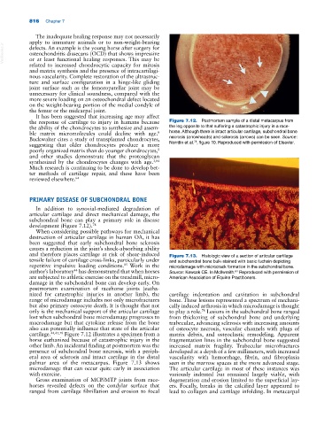
the response of cartilage to injury in humans because Figure 7.12. Postmortem sample of a distal metacarpus from
the ability of the chondrocytes to synthesize and assem the leg opposite to that suffering a catastrophic injury in a race-
ble matrix micromolecules could decline with age. horse. Although there is intact articular cartilage, subchondral bone
8
Buckwalter cites a study of transplanted chondrocytes, necrosis (arrowheads) and sclerosis (arrows) can be seen. Source:
78
suggesting that older chondrocytes produce a more Norrdin et al. , figure 10. Reproduced with permission of Elsevier.
poorly organized matrix than do younger chondrocytes,
8
and other studies demonstrate that the proteoglycan
synthesized by the chondrocytes changes with age. 3,84
Much research is continuing to be done to develop bet
ter methods of cartilage repair, and these have been
reviewed elsewhere. 64
PRIMARY DISEASE OF SUBCHONDRAL BONE
In addition to synovial‐mediated degradation of
articular cartilage and direct mechanical damage, the
subchondral bone can play a primary role in disease
development (Figure 7.12). 78
When considering possible pathways for mechanical
destruction of articular cartilage in human OA, it has
been suggested that early subchondral bone sclerosis
causes a reduction in the joint’s shock‐absorbing ability
and therefore places cartilage at risk of shear‐induced Figure 7.13. Histologic view of a section of articular cartilage
tensile failure of cartilage cross‐links, particularly under and subchondral bone bulk‐stained with basic fuchsin depicting
85
repetitive impulsive loading conditions. Work in the microdamage with microcrack formation in the subchondral bone.
author’s laboratory has demonstrated that when horses Source: Kawcak CE. In McIlwraith. Reproduced with permission of
44
64
are subjected to athletic exercise on the treadmill, micro American Association of Equine Practitioners.
damage in the subchondral bone can develop early. On
postmortem examination of racehorse joints (eutha
nized for catastrophic injuries in another limb), the cartilage indentation and cavitation in subchondral
range of microdamage includes not only microfractures bone. These lesions represented a spectrum of mechani
but also primary osteocyte death. It is thought that not cally induced arthrosis in which microdamage is thought
only is the mechanical support of the articular cartilage to play a role. Lesions in the subchondral bone ranged
78
lost when subchondral bone microdamage progresses to from thickening of subchondral bone and underlying
macrodamage but that cytokine release from the bone trabeculae, advancing sclerosis with increasing amounts
also can potentially influence that state of the articular of osteocyte necrosis, vascular channels with plugs of
cartilage. 44,45,78 Figure 7.12 illustrates a specimen from a matrix debris, and osteoclastic remodeling. Apparent
horse euthanized because of catastrophic injury in the fragmentation lines in the subchondral bone suggested
other limb. An incidental finding at postmortem was the increased matrix fragility. Trabecular microfractures
presence of subchondral bone necrosis, with a periph developed at a depth of a few millimeters, with increased
eral area of sclerosis and intact cartilage in the distal vascularity with hemorrhage, fibrin, and fibroplasia
palmar area of the metacarpus. Figure 7.13 shows seen in the marrow spaces at the more advanced stage.
microdamage that can occur quite early in association The articular cartilage in most of these instances was
with exercise. variously indented but remained largely viable, with
Gross examination of MCP/MTP joints from race degeneration and erosion limited to the superficial lay
horses revealed defects on the condylar surface that ers. Focally, breaks in the calcified layer appeared to
ranged from cartilage fibrillation and erosion to focal lead to collagen and cartilage infolding. In metacarpal