
Page 855 - Adams and Stashak's Lameness in Horses, 7th Edition
P. 855
Principles of Musculoskeletal Disease 821
domesticated breeding practices. In addition, breeding leading to chondronecrosis in the articular–epiphyseal
122
132
for rapidly growing offspring that hopefully will do bet cartilage complex, causing OCD‐type lesions. Lecocq
VetBooks.ir and protein appear to predispose these animals to DOD fetal and newborn foals at common sites of DOD and
ter in the show ring and feeding rations high in energy
et al. demonstrated the histologic changes that occur in
concluded that abnormalities in proteoglycan and colla
abnormalities presumably by contributing to rapid bone
growth. Other risk factors include mineral imbalances gen metabolism, cartilage canals, and vascularization of
such as copper deficiency or excess zinc, trauma, and the epiphyseal growth cartilage play a role in DOD.
55
genetic predilection. 99,125 Trace mineral deficiencies, cop However, in most cases, the underlying cause of the DOD
per in particular, have been incriminated in physitis and condition is multifactorial, usually obscure, and often
105
angular deformities in cattle, and copper has been never determined. (The reader is referred to Chapter 10
131
shown to cause clinical signs and joint pathology con for further information on specific DOD conditions such
sistent with osteochondrosis in foals. 43,44 Copper is as physitis and ALDs.)
required for the enzyme lysyl oxidase, which itself is
necessary for collagen cross‐linking. Therefore, defective Incomplete Cuboidal Bone Ossification/Juvenile Spavin
collagen cross‐linking may impair the strength of bone
collagen, producing essentially a “soft bone syndrome” Incomplete ossification of the cuboidal bones of the
particularly in the metaphyseal regions. Excess zinc or carpus or tarsus occurs most commonly in premature,
44
alterations in the calcium/phosphorus ratios in the diet twins, or underdeveloped newborn foals. At birth, the
may also lead to clinical problems of DOD, but these are cuboidal bones have not ossified sufficiently to with
less well defined in horses than copper deficiency. 99 stand the forces of normal weight‐bearing, predisposing
Trauma to the metaphyseal or epiphyseal growth to variable degrees of carpal or tarsal bone wedging or
plates can contribute to altered growth, subchondral collapse (Figure 7.15). Incomplete ossification without
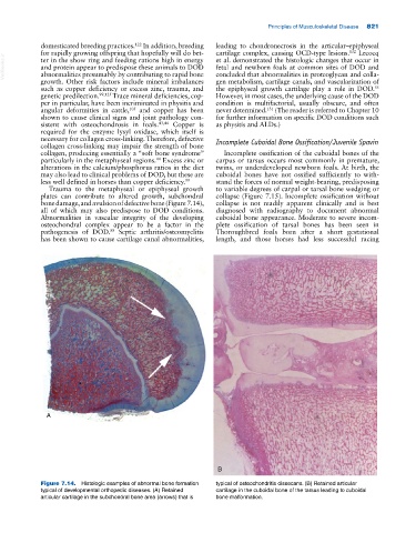
bone damage, and avulsion of defective bone (Figure 7.14), collapse is not readily apparent clinically and is best
all of which may also predispose to DOD conditions. diagnosed with radiography to document abnormal
Abnormalities in vascular integrity of the developing cuboidal bone appearance. Moderate to severe incom
osteochondral complex appear to be a factor in the plete ossification of tarsal bones has been seen in
pathogenesis of DOD. Septic arthritis/osteomyelitis Thoroughbred foals born after a short gestational
88
has been shown to cause cartilage canal abnormalities, length, and those horses had less successful racing
A
B
Figure 7.14. Histologic examples of abnormal bone formation typical of osteochondritis dissecans. (B) Retained articular
typical of developmental orthopedic diseases. (A) Retained cartilage in the cuboidal bone of the tarsus leading to cuboidal
articular cartilage in the subchondral bone area (arrows) that is bone malformation.