
Page 856 - Adams and Stashak's Lameness in Horses, 7th Edition
P. 856
822 Chapter 7
VetBooks.ir
A B C
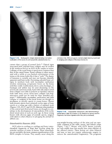
Figure 7.15. Radiographic images demonstrating incomplete premature foal. Without support, normal weight‐bearing could lead
ossification of the tarsal (A) and carpal (B) cuboidal bones in a to wedging and collapse of the tarsal bones (C).
careers than a group of normal foals. Clinical signs
38
associated with cuboidal bone collapse may be evident
in the newborn foal as an ALD of the carpus or tarsus.
Collapse of the tarsal bones is much more common than
that of the carpal bones. Tarsal collapse is often associ
ated with a sickle or cow‐hocked conformation of the
tarsus or the tarsus looks like it has a “curb.” The degree
of lameness is variable and may not become clinically
apparent until later in life. Preventing cuboidal bone col
lapse in newborn foals with incomplete ossification
involves minimizing compressive forces on the bones
until they ossify. Confinement, sleeve casts, bandages, or
bandages and splints may be used depending on the
severity. In a previous study, foals with only minor tarsal
bone collapse were able to perform as intended, whereas
foals with more severe tarsal bone collapse and frag
mentation could not be used for their intended pur
23
poses. Additionally, incomplete ossification and mild
collapse or wedging of the tarsal bones are thought to
predispose to juvenile spavin in young horses. Horses
with juvenile spavin have relatively severe signs of bone
spavin at a young age with a history of minimal work
(Figures 5.81, 10.27, and 10.58). Inherent abnormalities
of the central and third tarsal bones are thought to con
tribute to the development of osteoarthritis (OA) in the Figure 7.16. Lateromedial radiographic view demonstrating a
distal tarsal joints at such an early age. In those young typical lateral ridge OCD lesion of the distal femur (arrow) and the
horses with obvious signs of juvenile spavin, surgical fragments that have migrated within the joint (arrowhead).
arthrodesis is often effective in salvaging the animal for
athletic use.
Osteochondritis Dissecans (OCD) non‐weight‐bearing surfaces of the joint and are espe
cially common in the stifle, tarsus, and fetlock joints
OCD refers to cartilage or cartilage and bone (osteo (Figure 7.16). Horses with OCD lesions typically
125
chondral) fragments or flaps that develop along the are only mildly lame but usually have joint effusion of
articular surfaces of joints in horses. These abnormali the affected joint(s). These lesions are often bilateral
ties are probably the most common manifestation of the and may or may not require arthroscopic surgery to
DOD complex in horses. They usually occur along the remove the osteochondral fragment(s). The prognosis