
Page 860 - Adams and Stashak's Lameness in Horses, 7th Edition
P. 860
826 Chapter 7
VetBooks.ir
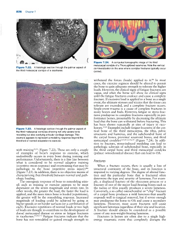
Figure 7.24. A computed tomographic image of the third
metacarpal condyle of a Thoroughbred racehorse. Note the vertical
Figure 7.22. A histologic section through the palmar aspect of demineralization in the area where condylar fractures typically occur
the third metacarpal condyle of a racehorse. (arrow).
withstand the forces (loads) applied to it. In most
84
cases, the exercise regimen should be altered to permit
the bone to gain adequate strength to tolerate the higher
loads. However, the clinical signs of fatigue fractures are
vague, and often the horse will show no clinical signs
until the fatigue fractures coalesce and cause a complete
fracture. If excessive load is applied to a bone as a single
event, the ultimate stresses and strains that the tissue can
tolerate are exceeded, and a complete fracture occurs.
Single‐event trauma is a cause of complete fractures in
many horses and foals. However, fatigue or stress frac
tures predispose to complete fractures especially in per
formance horses, presumably by decreasing the ultimate
load that the bone can withstand before fracturing. This
has been shown repeatedly at sites of injury in race
horses. 19,52 Examples include fatigue fractures of the cor
Figure 7.23. A histologic section through the palmar aspect of
the third metacarpal condyles showing not only severe bone tical bone of the third metacarpus, the tibia, pelvic
modeling but also overlying articular cartilage damage. This structures and humerus, and the subchondral bone of
example appears to represent a modeling response beyond the the carpal bones, proximal sesamoid bones, and third
threshold of normal adaptation to exercise. metacarpal condyles 5,65,115,118,130 (Figure 7.24). In addi
tion to fracture, stress‐induced modeling can lead to
pathologic sclerosis of subchondral bone, especially in
with training 19,52 (Figure 7.22). These are only a couple the third carpal bone and third metacarpal condyles
of examples of bone’s response to exercise, which (palmar osteochondral disease) that can lead to OA.
undoubtedly occurs in every bone during training and
performance. Unfortunately, there is a fine line between
what is considered to be normal adaptive training Fractures
(repetitive stress response) and overtraining that may be When a fracture occurs, there is usually a loss of
106
pathologic to the bone (repetitive stress injury) structural continuity of the bone, and its function is
(Figure 7.23). In addition, there is no objective means of impaired to varying degrees. The degree of altered func
characterizing that threshold between normal and path tion and the particular bone that is fractured often
ologic loading. determine the type and severity of lameness. For exam
The osteogenic response of bone to remodeling stim ple, a displaced fracture of the olecranon process or a
uli such as training or exercise appears to be most fracture of one of the major load‐bearing bones such as
dependent on the strain magnitude and strain rate. In the radius or tibia usually produces a severe lameness.
other words, the greater the load, the faster the load is Conversely, a so‐called osteochondral or “chip” fracture
applied, and the more times bone is loaded (repetitions), of a carpal bone produces a mild lameness that usually
the greater will be the remodeling response. Increased responds well to surgical removal, but if severe enough
magnitude of loading could be achieved by going at may predispose the horse to OA and cause a secondary
higher speeds or on harder surfaces (or a combination of lameness. However, most acute fractures will cause
each). Excessive repetitions of normal loads or too high significant lameness regardless of their size and location.
of loads are thought to contribute to the development of A fracture should always be considered as a possible
dorsal metacarpal disease or stress or fatigue fractures cause of any non‐weight‐bearing lameness.
in racehorses. 83,84,114 Fatigue fractures indicate that the Fractures in horses are often due to a single high‐
bone has not remodeled or gained enough strength to energy traumatic event that completely disrupts the