
Page 862 - Adams and Stashak's Lameness in Horses, 7th Edition
P. 862
828 Chapter 7
upper (humerus, ulna, and femur) aspects of the horse’s
limbs rarely become open, suggesting that proper immo
VetBooks.ir tarsus, radius, and tibia is the most critical to prevent the
bilization of fractures involving the metacarpus/meta
development of an open fracture during transport.
Fractures at these sites often require emergency surgical
repair to prevent open fracture configuration. The
reader is referred to the section on musculoskeletal
emergencies in Chapter 12 for specific details on frac
ture immobilization.
Fracture Healing in Horses
Fracture healing can be considered a series of pro
cesses that occur in sequence but are often overlapping.
The healing process can be divided into three distinct
phases: inflammatory, reparative, and remodeling.
63
During this process the bone will unite by one of two
patterns: primary or direct healing and secondary or
indirect healing. With primary bone healing, the bone
ends heal directly by Haversian remodeling in contact
and noncontact areas without the formation of a bone
callus. Rigid fracture stabilization and correct anatomi
cal reduction of the fracture are required for primary or
direct bone healing to occur. With indirect or secondary
bone healing, fibrous tissue or fibrocartilage is formed
initially between the fracture fragments with subsequent
replacement with new bone. Periosteal and endosteal cal
lus is formed to unite the bone ends. In horses, because
63
of their large size and weight, primary healing is rare,
and even with rigid internal fixation, a combination of
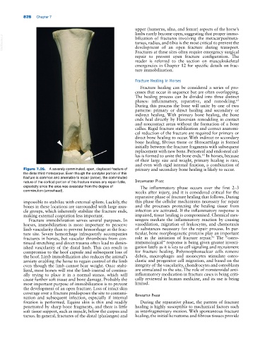
Figure 7.26. A severely comminuted, open, displaced fracture of primary and secondary bone healing is likely to occur.
the distal third metacarpus. Even though the condylar portion of this
fracture is common and amenable to repair (arrow), the comminuted
nature of the cortical portion of this fracture makes any repair futile, Inflammatory Phase
especially since the area was avascular from the degree of The inflammatory phase occurs over the first 2–3
comminution (arrowhead). weeks after injury, and it is considered critical for the
reparative phase of fracture healing that follows. During
impossible to stabilize with external splints. Luckily, the this phase the cellular mechanisms necessary for repair
bones in these locations are surrounded with large mus and the processes protecting the healing tissue from
cle groups, which inherently stabilize the fracture ends, infection are activated. If the inflammatory response is
making external coaptation less important. impaired, tissue healing is compromised. Chemical mes
Fracture immobilization serves several purposes. In sengers mediate the inflammatory reaction by causing
horses, immobilization is more important to preserve vasodilation, migration of leukocytes, and chemotaxis
limb vascularity than to prevent hemorrhage at the frac of substances necessary for the repair process. In par
ture site. Severe hemorrhage infrequently accompanies ticular, bone morphogenetic proteins play an important
91
fractures in horses, but vascular thrombosis from con role in the initiation of fracture repair. The “osteo‐
tinued stretching and direct trauma often lead to dimin immunological” response is being given greater investi
ished vascularity of the distal limb. This can result in gation lately as it is key to cell signaling and recruitment
compromise to the hoof capsule and subsequent loss of for fracture healing. Polymorphonuclear cells remove
the hoof. Limb immobilization also reduces the animal’s debris, macrophages and monocytes stimulate osteo
anxiety enabling the horse to regain control of the limb clastic and progenitor cell migration, and based on the
even though the limb cannot bear weight. Once stabi integrity of the vascularity, chondrocytes and osteoblasts
lized, most horses will rest the limb instead of continu are stimulated to the site. The role of nonsteroidal anti‐
ally trying to place it in a normal stance, which will inflammatory medication in fracture cases is being criti
cause further soft tissue and bone damage. Probably the cally reviewed in human medicine, and its use is being
most important purpose of immobilization is to prevent limited.
the development of an open fracture. Loss of intact skin
coverage over a fracture predisposes the site to contami
nation and subsequent infection, especially if internal reParatIve Phase
fixation is performed. Equine skin is thin and readily During the reparative phase, the pattern of fracture
penetrated by sharp bone fragments, and there is little healing is highly susceptible to mechanical factors such
soft tissue support, such as muscle, below the carpus and as interfragmentary motion. With spontaneous fracture
tarsus. In general, fractures of the distal (phalanges) and healing, the initial hematoma and fibrous tissues provide