
Page 857 - Adams and Stashak's Lameness in Horses, 7th Edition
P. 857
Principles of Musculoskeletal Disease 823
for performance of horses with OCD lesions is usually that contribute to clinical problems involve the stifle,
very good. More detailed discussion of OCD lesions can fetlock, pastern, coffin, and elbow joints (Figure 7.17).
VetBooks.ir caused by a defect in endochondral ossification, intra‐
Controversy exists as to whether these lesions are
be found in Chapter 10.
articular subchondral bone trauma, or a combination
Subchondral Cystic Lesions (SCLs)
of both. 6,12,47 Joint trauma can lead to the develop
SCLs (bone cysts or osseous cyst‐like lesions) are com ment of SCLs, and this has been shown experimentally
monly recognized pathologic entities of bones and joints and has been seen clinically (Figure 7.18). However,
6
in horses that may or may not cause lameness. SCLs may many of these lesions are seen in young horses and
be nonarticular or articular. However, most lesions that are bilateral, suggesting a developmental defect. SCLs
contribute to lameness involve the weight‐bearing area of
an articular surface. Nonarticular lesions (which may or
may not be classified as SCLs) usually involve the meta
physis and can go undiagnosed because they may not
cause clinical signs and normal bone remodeling may
resolve the defect. The most common age for diagnosis of
SCLs, or at least the time when clinical signs develop, is
usually 3 years or less. 6,42 However, horses demonstrate
clinical signs related to SCLs over a wide age range, and
the relationship between when the lesion develops and
when the horse begins to show clinical signs is not
known. This relationship probably varies depending on
47
the specific site of the SCL (most common location is the
medial femoral condyle), the age when the lesion devel
ops, and the occupation of the horse. Although most cases
of SCL have a developmental cause, older horses with OA
can develop SCL likely due to trauma. What causes or
initiates the appearance of clinical signs in horses with
articular SCLs remains unknown; however, the consistent
morphologic characteristics of the lesions suggest a com
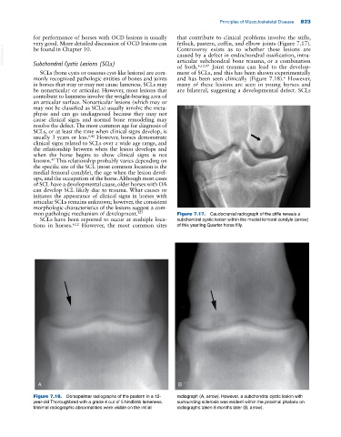
mon pathologic mechanism of development. 123 Figure 7.17. Caudocranial radiograph of the stifle reveals a
SCLs have been reported to occur at multiple loca subchondral cystic lesion within the medial femoral condyle (arrow)
tions in horses. 6,12 However, the most common sites of this yearling Quarter horse filly.
A B
Figure 7.18. Dorsopalmar radiographs of the pastern in a 12‐ radiograph (A, arrow). However, a subchondral cystic lesion with
year‐old Thoroughbred with a grade 4 out of 5 hindlimb lameness. surrounding sclerosis was evident within the proximal phalanx on
Minimal radiographic abnormalities were visible on the initial radiographs taken 6 months later (B, arrow).