
Page 861 - Adams and Stashak's Lameness in Horses, 7th Edition
P. 861
Principles of Musculoskeletal Disease 827
bone (breaks the bone into at least two pieces). In contrast
to most complete fractures, fatigue fractures are due to
VetBooks.ir single traumatic event that fractures the bone.
chronic trauma that weakens the bone, rather than a
114
Specifically, chronic repetitive stress normally induces
a bone strengthening cascade as a form of adaptation to
withstand these forces (repetitive stress response). This
is a normal process in all athletes, especially the horse,
and creates normal patterns of bone enlargement or
thickening, such as sclerosis through the processes of
bone modeling and remodeling. Specifically, repetitive
stress can induce the response one of two ways.
Microdamage can stimulate remodeling events and lead
to replacement of the damaged bone. Since osteoclastic
function is more rapid than the osteoblastic response,
this does create a window of vulnerability in which fur
ther high stresses can induce more damage within the
relatively porous bone. The second form of bone
strengthening in response to stress is through bone mod
eling, in which osteoid and mineral are added to existing
bone to create either a dense sclerotic region (such as in
subchondral bone) or geometric changes to bone archi
tecture in order to maximize stress resistance (such as in
cortical bone). This modeled bone is often more brittle
than normal and in some instances can create pain and
necrosis in these areas.
Fatigue fractures (repetitive stress injury) of the
humerus, tibia, pelvis, and metacarpus/metatarsus
(Figure 7.25) are commonly diagnosed in performance
horses (primarily racehorses). 114,115 Initially these lesions
cause variable degrees of lameness but may contribute
to complete fracture of the involved bone with contin
ued use. Diagnosis of incomplete fractures in race
114
horses is critical to prevent catastrophic bone failure,
but this can be difficult and the timing is critical. These
horses may not be overtly lame at the time of examina
tion, but may have a history of not working correctly, or
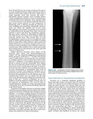
be lame intermittently or immediately after work. If Figure 7.25. A radiograph of the third metacarpal bone from a
lameness is present, then diagnostic analgesia can be 2‐year‐old racing Quarter Horse showing a fatigue fracture of the
performed, but the proximal location of many of these dorsal aspect of the bone (arrows).
fractures often precludes its use. In addition, many sub
chondral bone lesions may not extend into the joint
early in the disease process, making intra‐articular anal Fracture Stabilization for Transport/Fracture Immobilization
21
gesia of little use. Nuclear scintigraphy can be very
helpful in locating suspected fatigue fractures that are Fractures are a frequently diagnosed problem in
not apparent on radiographs. Care must be taken in horses that often require emergency first aid treatment
overinterpretation though, as young exercising horses (Table 12.1). Horses are not readily ambulatory on three
will typically show sites of intense remodeling, especially limbs and often become very anxious when they are
in the fetlock joints. unable to place weight on a fractured limb, which poten
Treatment of incomplete fractures is much less compli tially can result in further injury. First aid measures
cated than treating complete fractures and usually involves should be directed toward minimizing further damage
a period of inactivity combined with a change in training to the fractured limb and maintaining it in a position
schedule. In contrast, complete fractures of long bones and condition that will facilitate repair. The goals of first
119
of horses are one of the most difficult injuries to treat suc aid fracture management are to prevent damage to neu
cessfully, and many horses are still euthanized because of ral and vascular elements of the limb, to prevent skin
the severity of the fracture (Figure 7.26). The prognosis of penetration of the fracture fragments or minimize fur
repairing complete fractures in horses depends on the spe ther contamination of an existing wound, to relieve anx
cific bone affected, the temperament of the horse, the age iety of the animal by stabilizing the fractured limb, to
and size of the horse, the specific characteristics of the prevent deterioration of the patient’s physiologic status,
fracture, and the expertise of the surgeon, to name just a and to minimize further damage to the fractured bone
few. However, proper stabilization of the fracture for ends and surrounding soft tissue. 11,13 Most of these
transport to a surgical facility for repair is crucial to objectives can be accomplished by proper stabilization
achieving a favorable outcome, and newer repair tech or splinting of the fracture. However, fractures of the
niques and implants have improved prognosis. upper forelimb and hindlimb in horses are nearly