
Page 859 - Adams and Stashak's Lameness in Horses, 7th Edition
P. 859
Principles of Musculoskeletal Disease 825
VetBooks.ir
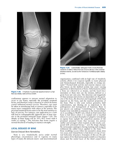
Figure 7.21. Lateromedial radiograph from a horse that was
casted for 6 weeks. Notice the loss of bone density in the proximal
sesamoid bones, as well as the formation of enthesophytes distally
(arrow).
organization, combined with its high rate of metabolic
activity, allows it to respond rapidly to many physical
and biomechanical demands, especially in young ani
mals. Normal daily activity and exercise impose com
62
plex forces on the skeletal system that cause varying
quantities of deformations of bone. Deformations within
a bone are referred to as strains, and the local force
Figure 7.20. A fracture of a proximal sesamoid bone in a foal intensities at these sites are the stresses, defined as a
that was initially stall confined at birth.
given force per unit area. The direction and magnitude
of these stresses and strains are dependent on the geom
confinement appears to prevent normal adaptation to etry of the bone, the direction and magnitude of the load
occur to the bones, especially the proximal sesamoid imposed on the bone, and the material properties of
bones, and therefore create a situation in which the bones the bone. That is why joint surface geometry, limb
cannot withstand normal exercise. Therefore, care must conformation, and shoeing are so important to the
be taken in introducing foals to turnout, especially if the pathogenesis and management of lameness. Bone in
mares sense competition with others in the pasture. The any species can undergo “remodeling” or “modeling.”
proximal sesamoid bones seem to be the most sensitive to Equine bone remodels in response to the stresses placed
lack of exercise. For instance, cast application to a limb on it according to Wolff’s law, which in brief states that
will lead to radiographically apparent loss of bone den bone is laid down where it is needed and is resorbed
sity in the proximal sesamoid bones (Figure 7.21). The where it is not needed. Examples of adaptive remodeling
density of these bones will be 30%–40% lower with 6 in equine bone include the cancellous portion of the
weeks of casting. This indicates that bones require a proximal sesamoid bones that becomes less porous
35
normal threshold of loading to maintain strength. (stiffer and stronger) when trained on dirt tracks com
pared with untrained horses, and the shape changes
133
the metacarpus undergoes during training and racing to
LOCAL DISEASES OF BONE withstand the added forces applied to the bone. 79,83,84
Exercise‐Induced Bone Remodeling Bone modeling occurs when the resorptive phase does
not occur, and instead bone is laid down on top of nor
Bone is very metabolically active under normal mal trabecular structures. Examples of this are the pal
physiologic circumstances and in response to many mar aspect of the third metacarpal condyle as it becomes
types of injury. Its intricate micro‐ and macrostructural dense and the radial facet of the third carpal bone