
Page 863 - Adams and Stashak's Lameness in Horses, 7th Edition
P. 863
Principles of Musculoskeletal Disease 829
some internal stress and support, and periosteal and Fracture Fixation
endosteal callus formation provides interfragmentary In horses, more than any other domestic animal one
VetBooks.ir nous and endochondral ossification. This process can has to carefully define “successful” fracture healing. For
stabilization, and bone union occurs by intramembra
63
centuries, bone has been observed to heal by production
take from 2 to 12 months to be completed depending on
the method of fracture fixation that was utilized, the sta of callus, but the end result was often angulation, rota
tion, or limb shortening. With intra‐articular fractures, a
bility of the fracture, and the size of the fracture gap certain amount of OA was often the end result. Fixation
(fracture displacement). techniques used to achieve primary bone healing with
intra‐articular fractures have greatly decreased the mor
remodelIng Phase bidity associated with OA in these cases. In addition,
improved techniques in internal fixation of long bone
The remodeling phase occurs during and following the
reparative phase. Avascular and necrotic regions of bone fractures have emphasized improving the implants to
withstand massive functional forces, thus preventing
are replaced by Haversian remodeling. Malalignment of failure due to mechanical overload. Such implants must
63
fracture fragments may be corrected during this phase of also be strong enough to maintain their integrity until
healing by remodeling of the fracture site and functional the bone has united, without breaking under fatigue.
adaptation, particularly in young animals. With weight‐ However, despite improvements in fracture fixation
bearing and loading of the fracture, bone is removed from equipment, anesthetic protocols, and recovery methods,
the convex surfaces and laid down on the concave sur successful repair of some long bone fractures in horses
faces. This process tends to realign the bone after malun remains very difficult.
ion (Figure 7.27). However, fracture remodeling cannot Stress protection is a phenomenon seen when a bone
correct torsional deformities associated with fracture that has been rigidly immobilized by a plate(s) undergoes
healing. Theoretically, bone can heal completely and certain histologic events, including loss of bone mass
regain pre‐fracture strength and function. without a corresponding reduction in size (quantitative
osteopenia). Stress protection results in Haversian
119
remodeling and has generated considerable interest in
man and small animals because of the potential for
refracture of the bone following removal of the plate.
Stress protection is almost an unknown occurrence in
the horse, even in foals, because of the greater loads
imparted on the implants compared to man and other
smaller animals. In fact, the size and weight of horses
119
often stress the limits of stress tolerance in implants.
While the emphasis of research activity in man and
small animals has focused on the development of more
flexible implants, in horses the emphasis has been in
the reverse direction to provide stronger implants in
an attempt to overcome the massive loading of the
implants.
An important consideration in the horse is stress
concentration. This is where biomechanical loads are
concentrated in a small area of normal or weakened
119
bone, potentially leading to complete bone failure.
This primarily occurs in the diaphysis of long bones but
may also develop elsewhere along the bone. Examples
of stress concentration include drill holes that are not
filled with implants during internal fixation and vacant
screw holes after implant removal (such as after
119
metacarpal stress fracture repair). Additionally, stress
concentration occurs at the ends of bone plates espe
cially if they stop in the mid‐diaphyseal region of a bone
and at intramedullary (IM) pin hole sites following
removal of external fixators. These locations are all
areas where small areas of cortical bone are absent or
have been weakened and can fail if excessive loading of
the bone occurs.
ComPressIon fIxatIon
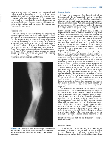
Figure 7.27. Radiograph of the third metacarpal bone of a horse The use of various methods of compression in the
that presented several months after the fracture had been treated treatment of fractures in man and animals is widely
with external splinting. The fracture had healed but was severely accepted. Under stable conditions it is recognized that
malaligned. both cancellous and cortical bones heal by primary bone