
Page 1203 - Equine Clinical Medicine, Surgery and Reproduction, 2nd Edition
P. 1203
1178 CHAPTER 11
VetBooks.ir heal (see Figs. 11.36, 11.37). Cyanoacrylate adhe- is greater than 50% of the corneal depth, stromal
necrosis (melting) is present or the ulcer is pro-
sive (tissue glue) may also be used in some cases of
corneal ulceration; however, it can be expensive, is
A lamellar keratectomy, in which the anterior stroma
infrequently indicated and application can be dif- gressing despite appropriate medical management.
ficult. It should not be used to seal leaking corneal and epithelium are surgically removed, is performed
perforations and is not recommended for use in des- to eliminate diseased tissue (necrotic stroma),
cemetocoeles, because perforation may result from shorten the course of the disease and acquire tissues
the resulting heat and contraction. The use of a for diagnostic testing (Fig. 11.86). The keratectomy
well-fitting hood with a hard eye-cup, cross-tying or site may be repaired with placement of a conjunctival
constant supervision may be required to prevent self- pedicle graft (CPG), free conjunctival island graft,
trauma to a painful eye or perforation of an eye with tarsoconjunctival graft, porcine small-intestinal
a severely compromised cornea. submucosa graft, amnion graft, corneoscleral or cor-
When treating horses with corneal ulceration, neoconjunctival transposition, or corneal autograft,
frequent re-evaluations are required to determine allograft or heterograft. These measures are aimed
how rapidly the ulcer is changing, and treatment at increasing the patient’s comfort and preserving
should be modified according to the response to ocular integrity. Porcine small-intestinal submucosa
therapy. In addition to the depth of the corneal ulcer, is a collagen-based material that is safe, relatively
the eye should be evaluated closely at each examina- inexpensive, commercially available in individual
tion for evidence of corneal melting. In general, if packages, easy to handle and resistant to anticolla-
goals of therapy are not met on target, adjustments genase activity.
to medications should be made accordingly and the With lesions deeper than 50% of the corneal
need for surgical intervention and referral ascer- depth, CPGs and corneoscleral or corneoconjunc-
tained. Sequential photographs or detailed drawings tival transpositions are recommended. CPGs pro-
can be very helpful in documenting changes. vide mechanical reinforcement of the cornea and
Unfortunately, surgery may be necessary in addi- facilitate healing by providing tectonic support,
tion to medical management, and in these cases providing fibrovascular tissue to fill the stromal
expedient referral to an ophthalmic specialist is defect and delivering a direct vascular supply to the
vital. Surgical intervention can help by decreasing lesion, including antiproteolytic and antimicrobial
the dose, length of time and frequency at which agents (Figs. 11.87, 11.88). Vascular grafts offer
medications are administered more quickly than significant antimicrobial and anticollagenase activ-
with medical management alone. In general, sur- ity, which helps control bacterial infections and cor-
gery is recommended in those cases where the ulcer neal liquefaction. They provide a cornea compatible
11.86
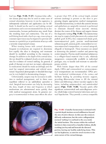
Fig. 11.86 A lamellar keratectomy is initiated with
a partial-depth incision several millimetres beyond
the area of active disease. In this case the cornea is
diffusely oedematous, but the active collagenolysis
(corneal ‘melting’) is confined to the area inside
the initial incision. The stroma within the outlined
incision is then removed in a lamellar fashion, ideally
reaching just beyond and beneath the diseased
portion, leaving any healthy underlying tissue. This
eye has hyphaema, an anterior uveal reaction to the
acute nature of the corneal damage.