
Page 1205 - Equine Clinical Medicine, Surgery and Reproduction, 2nd Edition
P. 1205
1180 CHAPTER 11
VetBooks.ir ventral conjunctival tissues are advanced centrally secondary to rapid enzymatic degradation of stromal
collagen and ground substance caused by infectious
and sutured together over the centre of the cornea.
It is typically used when the entire cornea is affected
mal abscessation in the horse can also progress to
with severe ulcerative keratitis and the availability and non-infectious ulcerative keratitis. Deep stro-
of microsurgical instruments and magnification is full-thickness corneal rupture in rare cases. Globe
limited; however, it should not be considered as a rupture may also occur during examination of deep
first-choice procedure. The main advantages of this corneal ulcers or descemetocoeles if the horse is not
technique are the relative speed with which the graft amenable to examination. Globe rupture results
can be harvested and closed, and the fact that actual in hyphaema, fibrin formation and uveal prolapse.
corneal suturing is not required. The major disad- Perforations may initially seal but are unstable and
vantages are the inability to view the eye while the may leak intermittently.
graft is in place and loss of the ability for the horse
to see through or around the graft during healing. Clinical presentation
Following resolution of the disease process the graft An iris prolapse typically appears as a focal red to
is opened and trimmed diligently to ensure minimal brown or tan corneal mass bulging from the surface
permanent scarring. and associated with corneal oedema and fibrin for-
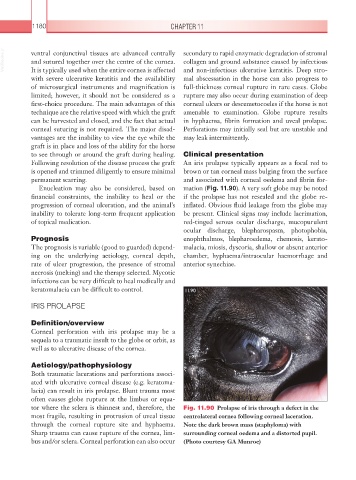
Enucleation may also be considered, based on mation (Fig. 11.90). A very soft globe may be noted
financial constraints, the inability to heal or the if the prolapse has not resealed and the globe re-
progression of corneal ulceration, and the animal’s inflated. Obvious fluid leakage from the globe may
inability to tolerate long-term frequent application be present. Clinical signs may include lacrimation,
of topical medication. red-tinged serous ocular discharge, mucopurulent
ocular discharge, blepharospasm, photophobia,
Prognosis enophthalmos, blepharoedema, chemosis, kerato-
The prognosis is variable (good to guarded) depend- malacia, miosis, dyscoria, shallow or absent anterior
ing on the underlying aetiology, corneal depth, chamber, hyphaema/intraocular haemorrhage and
rate of ulcer progression, the presence of stromal anterior synechiae.
necrosis (melting) and the therapy selected. Mycotic
infections can be very difficult to heal medically and
keratomalacia can be difficult to control. 11.90
IRIS PROLAPSE
Definition/overview
Corneal perforation with iris prolapse may be a
sequela to a traumatic insult to the globe or orbit, as
well as to ulcerative disease of the cornea.
Aetiology/pathophysiology
Both traumatic lacerations and perforations associ-
ated with ulcerative corneal disease (e.g. keratoma-
lacia) can result in iris prolapse. Blunt trauma most
often causes globe rupture at the limbus or equa-
tor where the sclera is thinnest and, therefore, the Fig. 11.90 Prolapse of iris through a defect in the
most fragile, resulting in protrusion of uveal tissue centrolateral cornea following corneal laceration.
through the corneal rupture site and hyphaema. Note the dark brown mass (staphyloma) with
Sharp trauma can cause rupture of the cornea, lim- surrounding corneal oedema and a distorted pupil.
bus and/or sclera. Corneal perforation can also occur (Photo courtesy GA Munroe)