
Page 1204 - Equine Clinical Medicine, Surgery and Reproduction, 2nd Edition
P. 1204
Eyes 1179
VetBooks.ir 11.87 11.88
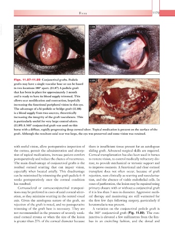
Figs. 11.87–11.89 Conjunctival grafts. Pedicle 11.89
grafts may have a single vascular base or can be based
in two locations 180° apart. (11.87) A pedicle graft
that has been in place for approximately 1 month
and is ready to have its blood supply trimmed. This
allows scar modification and contraction, hopefully
increasing the functional peripheral vision in this eye.
The advantage of a bi-pedicle or bridge graft (11.88)
is a blood supply from two sources, theoretically
increasing the integrity of the graft vasculature. This
is particularly useful for very large central ulcers.
(11.89) A 360° conjunctival graft was used on this
horse with a diffuse, rapidly progressing deep corneal ulcer. Topical medication is present on the surface of the
graft. Although the resultant axial scar was large, the eye was preserved and some vision was retained.
with useful vision, allow postoperative inspection of there is insufficient tissue present for an autologous
the cornea, permit the administration and absorp- sliding graft. Advanced surgical skills are required.
tion of topical medications, increase patient comfort Corneal transplantation has also been used in horses
postoperatively and reduce the chance of recurrence. to restore vision, to control medically refractory dis-
The main disadvantage of conjunctival grafts is the ease, to provide mechanical or tectonic support and
residual corneal scarring that can impair vision, to improve cosmesis. A functional and clear corneal
especially when located axially. This disadvantage transplant does not often occur, because of graft
can be minimised by trimming the graft pedicle 6–8 rejection, seen clinically as scarring and vascularisa-
weeks postoperatively once the corneal condition tion, and the absence of viable endothelial cells. In
has resolved. cases of perforation, the lesion may be repaired using
Corneoscleral or corneoconjunctival transposi- primary closure with or without a conjunctival graft
tions may be preferred in cases of axial corneal ulcer- if it is less than 3 mm in diameter. Aggressive medi-
ation, as they minimise scarring in the central visual cal therapy and monitoring are still warranted for
axis. Given the autologous nature of the graft, no the first few days following surgery, particularly if
rejection of the graft is noted, and no postoperative keratomalacia was present.
trimming of the graft base is necessary. They are A variation on the conjunctival pedicle graft is
not recommended in the presence of severely weak- the 360° conjunctival graft (Fig. 11.89). The con-
ened corneal stroma or when the size of the lesion junctiva is elevated a few millimetres from the lim-
is greater than 25% of the corneal diameter because bus in an encircling fashion, and the dorsal and