Page 151 - Equine Clinical Medicine, Surgery and Reproduction, 2nd Edition
P. 151

126                                        CHAPTER 1


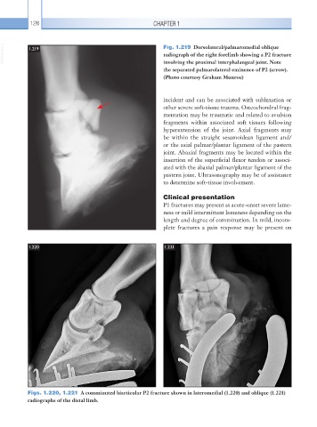
  VetBooks.ir  1.219                                      Fig. 1.219  Dorsolateral/palmaromedial oblique
                                                          radiograph of the right forelimb showing a P2 fracture
                                                          involving the proximal interphalangeal joint. Note
                                                          the separated palmarolateral eminence of P2 (arrow).
                                                          (Photo courtesy Graham Munroe)


                                                          incident and can be associated with subluxation or
                                                          other severe soft-tissue trauma. Osteochondral frag-
                                                          mentation may be traumatic and related to avulsion
                                                          fragments within associated soft tissues following
                                                          hyperextension of the joint. Axial fragments may
                                                          be within the straight sesamoidean ligament and/
                                                          or the axial palmar/plantar ligament of the pastern
                                                          joint. Abaxial fragments may be located within the
                                                          insertion of the superficial flexor tendon or associ-
                                                          ated with the abaxial palmar/plantar ligament of the
                                                          pastern joint. Ultrasonography may be of assistance
                                                          to determine soft-tissue involvement.

                                                          Clinical presentation
                                                          P1 fractures may present as acute-onset severe lame-
                                                          ness or mild intermittent lameness depending on the
                                                          length and degree of comminution. In mild, incom-
                                                          plete fractures a pain response may be present on


           1.220                                          1.221


           Figs. 1.220, 1.221  A comminuted biarticular P2 fracture shown in lateromedial (1.220) and oblique (1.221)
           radiographs of the distal limb.
   146   147   148   149   150   151   152   153   154   155   156