
Page 147 - Equine Clinical Medicine, Surgery and Reproduction, 2nd Edition
P. 147
122 CHAPTER 1
VetBooks.ir Tendon damage is seen as focal signal increase 4 months. Horses should be shod with egg-bar shoes
or back-to-front shoes, thereby reducing the oppor-
on both T1- and T2-weighted sequences, vari-
ably accompanied by enlargement of the affected
with heel elevation may be useful in horses with
lobe. There is a good correlation between the MRI tunity for hyperextension of the DIP joint. Shoeing
appearance and the pathological classification of severe lameness to improve lameness quickly, but it
lesions. Most tendon lesions are seen in the proximal has been suggested this should not be maintained for
recess of the navicular bursa. Lesions may be asso- longer than 3 months to avoid permanent functional
ciated with herniation of torn fibres and granuloma shortening of the DDFT during healing by fibrosis.
formation into the navicular bursa. Navicular bursi- In some horses, heel elevation exacerbates lameness.
tis may accompany the injury, with possible adhesion The outcome for horses with primary tendinitis
formation between the dorsal surface of the tendon treated with 6 months’ rest alone is disappointing,
and the collateral sesamoidean and impar ligaments. with only 25–30% returning to full athletic function
The latter phases of healing are by fibrosis and the and more than 60% suffering persistent or recurrent
healing rate can be followed on MRI scans. On CT lameness. Several additional therapies have been
images, tendon lesions may show focal hypoattenu- tried, including injection of corticosteroids into the
ation, enlargement of a tendon lobe and contrast navicular bursa and/or the digital sheath, shock-
enhancement in acute injuries. wave therapy, intralesional injection of biological
and regenerative products, inferior check ligament
Management desmotomy and bursoscopic debridement of dorsal
The most important aspect of treatment is a long surface tendon tears and granulomas (Figs. 1.212,
period of rest (6 months or more). This should con- 1.213). So far, only bursoscopic debridement and
sist of stall confinement with a low-grade mainte- intrabursal/intrathecal corticosteroid therapy have
nance daily exercise programme of 10–15 minutes been evaluated critically. In a study of 92 horses with
walking, starting after the first or second month intrabursal tears, 42% were able to return to their
of strict stall rest, depending on the severity of the original level of performance following bursoscopic
injury. Walking exercise can be increased gradu- debridement of surface lesions of the intrabursal
ally in duration but not intensity over the ensuing portion of the DDFT, but 37% of horses suffered
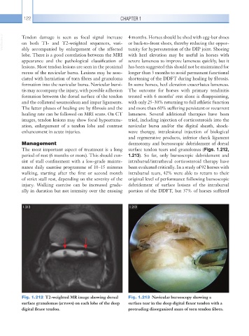
1.212 1.213
Fig. 1.212 T2-weighted MR image showing dorsal Fig. 1.213 Navicular bursoscopy showing a
surface granulomas (arrows) on each lobe of the deep surface tear in the deep digital flexor tendon with a
digital flexor tendon. protruding disorganised mass of torn tendon fibres.