
Page 143 - Equine Clinical Medicine, Surgery and Reproduction, 2nd Edition
P. 143
118 CHAPTER 1
VetBooks.ir 1.203 1.204
Figs. 1.203, 1.204 Septic navicular bursa. Radiographs showing the use of a solid probe (1.203) and liquid
contrast medium (1.204) to confirm communication between a wound in the ground surface of the foot and the
navicular bursa.
1.205 Management
Treatment of navicular bursal sepsis is always an
emergency and cases should be referred to a special-
ist facility to be further assessed. The horse is placed
on systemic antibiotics and NSAIDs, and tetanus
prophylaxis provided. Most horses that present with
severe lameness, typically of several days’ duration,
require surgical intervention. The hoof capsule sur-
rounding the entry wound on the ground surface of
the foot should be removed and the tract exposed
to permit introduction of arthroscopic instruments
into the bursa. Bursoscopy, under general anaes-
thesia, is generally recommended because it enables
visualisation of the bursa including the entry wound
through the DDFT and damage to the navicular
bone, debridement of both the entry wound and the
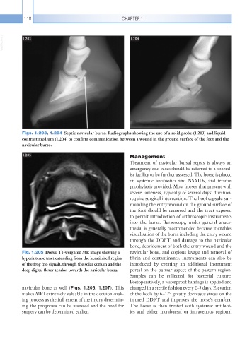
Fig. 1.205 Dorsal T1-weighted MR image showing a navicular bone, and copious lavage and removal of
hyperintense tract extending from the keratinised region fibrin and contaminants. Instruments can also be
of the frog (no signal), through the solar corium and the introduced by creating an additional instrument
deep digital flexor tendon towards the navicular bursa. portal on the palmar aspect of the pastern region.
Samples can be collected for bacterial culture.
Postoperatively, a waterproof bandage is applied and
navicular bone as well (Figs. 1.206, 1.207). This changed in a sterile fashion every 2–3 days. Elevation
makes MRI extremely valuable in the decision-mak- of the heels by 6–12° greatly decreases stress on the
ing process as the full extent of the injury determin- injured DDFT and improves the horse’s comfort.
ing the prognosis can be assessed and the need for The horse is then treated with systemic antibiot-
surgery can be determined earlier. ics and either intrabursal or intravenous regional