
Page 140 - Equine Clinical Medicine, Surgery and Reproduction, 2nd Edition
P. 140
Musculoskeletal system: 1.3 The foot 115
VetBooks.ir 1.200 1.201
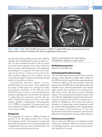
Figs. 1.200, 1.201 Dorsal (1.200) and transverse (1.201) T2-weighed MR images showing signal increase
characteristic of collateral desmitis of the distal interphalangeal joint (arrows).
into the soft working surfaces on that side, while the SEPTIC ARTHRITIS OF THE DISTAL
opposite shoe branch should be narrower than nor- INTERPHALANGEAL (DIP) JOINT
mal. The shoe should be bevelled in the toe region
and in the branch opposite to the injury. When the Definition/overview
injury is severe, with discontinuity of the ligament, Infection of the DIP joint.
immobilisation in a pastern–foot limb cast is indi-
cated for the first 4–6 weeks of the period of rest. Aetiology/pathophysiology
Daily in-hand walking for 10–15 minutes can be The DIP joint may become infected from traumatic
started after the first 2 months of stall rest but should injuries or joint injection; in foals, septic arthritis of
avoid circling and horse walkers. the DIP joint may be caused by haematogenous spread
Extracorporeal shock-wave therapy is recom- of infection. Injuries to the ground surface of the foot
mended during the rest period. Three applications that penetrate the DIP joint are also likely to affect the
are spaced 2 weeks apart over a period of 4 weeks. navicular bursa. Neurectomised horses may develop
Intra-articular medication of the DIP joint with septic arthritis from expansion of an unnoticed sub-
ACS/IRAP may be useful as an anti-inflammatory solar abscess. Joints that become infected following
substance diffusing from the joint into the ligament trauma to the coronary band and adjacent pastern may
or suppressing any concurrent synovitis and cap- also infect the digital flexor tendon sheath and navicu-
sulitis. Intralesional injection has been used under lar bursa. Iatrogenic joint infection is caused most fre-
imaging guidance (radiography, ultrasonography, quently by Staphylococcus aureus while joint infection
CT or MRI) for deposition of mesenchymal stem secondary to a penetrating wound is generally caused
cells or platelet-rich plasma (PRP), but their efficacy by a mixed bacterial population with a predominance
remains questionable. of gram-negative Enterobacteriaceae. It has been
shown that joint infections caused by S. aureus are more
Prognosis difficult to treat than those caused by other organisms.
The prognosis for return to athletic function is
guarded to fair with approximately 50% of horses Clinical presentation
recovering successfully. The prognosis is not adversely Horses with septic DIP joints are usually presented
affected by the presence of osseous abnormalities but with a severe lameness. There may also be evidence
is significantly worse for horses with instability, OA of swelling proximal to the coronary band, particu-
or other concurrent injuries in the foot. larly dorsally, or a wound.