
Page 141 - Equine Clinical Medicine, Surgery and Reproduction, 2nd Edition
P. 141
116 CHAPTER 1
VetBooks.ir Differential diagnosis and more accurate diagnosis of septic arthritis in
horses as compared with other imaging modalities,
Abscess; fracture of the distal phalanx or navicular
bone; severe strain or sprain; other deep digital sepsis.
especially when the clinical diagnosis is challenging.
Diagnosis Management
Sepsis should be strongly considered when a horse Treatment of a septic DIP joint involves aggressive
presents with a severe lameness and diffuse swelling arthroscopic joint lavage, systemic broad-spectrum
proximal to the coronary band dorsally and is pain- antibiotics, regional infusion of antibiotics, intrasy-
ful on flexion of the digit. A history of a recent injec- novial antibiotics and NSAIDs. Samples should be
tion into the joint, or the presence of a wound in the collected for bacterial culture. Arthroscopic lavage
dorsal to middle third of the frog or proximal to the and debridement should be performed via both
coronary band adjacent to the joint capsule, should the dorsal and palmar/plantar pouches of the joint
raise the index of suspicion. Confirmation of sepsis to remove intra-articular pannus effectively. An
is achieved by centesis (elevated WBC count and the ingress drain can be placed in the dorsal pouch of
identification of bacteria) or by confirming commu- the DIP joint to facilitate continuous or repeated
nication of the joint cavity with an external wound. intra-articular administration of antimicrobials.
Exploration of wounds proximal to the coronary Antimicrobials should be continued for at least
band, either digitally or with a sterile probe, may 2 weeks after closure of any wound communication
readily demonstrate joint involvement. Infusion of with the joint, the lameness has resolved or after the
sterile saline into the joint from a site remote to the DIP joint synovial fluid analysis has returned to nor-
wound, and observing it exiting a wound, confirms mal. In the presence of chronic articular cartilage
the communication definitively. Radiography is not or subchondral bone loss, or if infection cannot be
usually helpful in the diagnosis of acute joint sepsis, successfully removed, ankylosis of the DIP joint can
but later on in the course of the disease loss of joint be attempted to obtain pasture (breeding) soundness
space, subchondral lysis and periarticular new bone with permanent mechanical lameness.
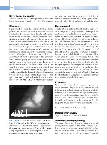
may be present (Fig. 1.202). MRI may allow earlier
Prognosis
Failure to eliminate infection results in permanent,
1.202 severe lameness, likely overload laminitis in the con-
tralateral limb and euthanasia. For horses in which the
infection can be controlled before significant degener-
ation of the articular surface occurs, the prognosis for
return to work is fair. For those horses in which there
is significant degeneration of the articular surface, the
long-term prognosis for survival is guarded to poor.
SEPTIC NAVICULAR BURSITIS
Definition/overview
Septic synovitis of the navicular bursa.
Fig. 1.202 Septic distal interphalangeal (DIP) joint. Aetiology/pathophysiology
Lateral radiograph of the DIP joint demonstrating The cause of a septic navicular bursa is almost invari-
extensive loss of the articular surfaces, exostoses on ably a puncture wound to the ground surface of the
the dorsal and palmar surfaces of the middle phalanx, foot in an area centred on the middle third of the frog
a large sequestrum of the extensor process of the and its collateral sulci. Such solar punctures have
distal phalanx and subluxation of the joint. been referred to historically as ‘streetnail’ injuries.