
Page 212 - Equine Clinical Medicine, Surgery and Reproduction, 2nd Edition
P. 212
Musculoskeletal system: 1.5 The hindlimb 187
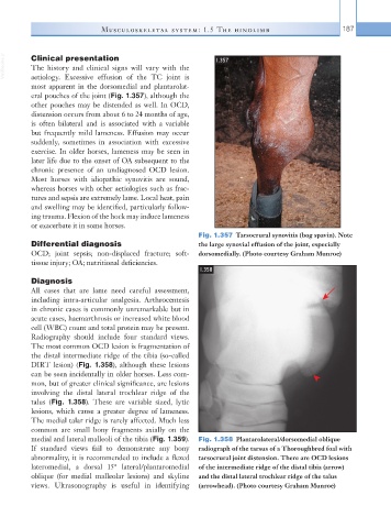
VetBooks.ir Clinical presentation 1.357
The history and clinical signs will vary with the
aetiology. Excessive effusion of the TC joint is
most apparent in the dorsomedial and plantarolat-
eral pouches of the joint (Fig. 1.357), although the
other pouches may be distended as well. In OCD,
distension occurs from about 6 to 24 months of age,
is often bilateral and is associated with a variable
but frequently mild lameness. Effusion may occur
suddenly, sometimes in association with excessive
exercise. In older horses, lameness may be seen in
later life due to the onset of OA subsequent to the
chronic presence of an undiagnosed OCD lesion.
Most horses with idiopathic synovitis are sound,
whereas horses with other aetiologies such as frac-
tures and sepsis are extremely lame. Local heat, pain
and swelling may be identified, particularly follow-
ing trauma. Flexion of the hock may induce lameness
or exacerbate it in some horses.
Fig. 1.357 Tarsocrural synovitis (bog spavin). Note
Differential diagnosis the large synovial effusion of the joint, especially
OCD; joint sepsis; non-displaced fracture; soft- dorsomedially. (Photo courtesy Graham Munroe)
tissue injury; OA; nutritional deficiencies.
1.358
Diagnosis
All cases that are lame need careful assessment,
including intra-articular analgesia. Arthrocentesis
in chronic cases is commonly unremarkable but in
acute cases, haemarthrosis or increased white blood
cell (WBC) count and total protein may be present.
Radiography should include four standard views.
The most common OCD lesion is fragmentation of
the distal intermediate ridge of the tibia (so-called
DIRT lesion) (Fig. 1.358), although these lesions
can be seen incidentally in older horses. Less com-
mon, but of greater clinical significance, are lesions
involving the distal lateral trochlear ridge of the
talus (Fig. 1.358). These are variable sized, lytic
lesions, which cause a greater degree of lameness.
The medial talar ridge is rarely affected. Much less
common are small bony fragments axially on the
medial and lateral malleoli of the tibia (Fig. 1.359). Fig. 1.358 Plantarolateral/dorsomedial oblique
If standard views fail to demonstrate any bony radiograph of the tarsus of a Thoroughbred foal with
abnormality, it is recommended to include a flexed tarsocrural joint distension. There are OCD lesions
lateromedial, a dorsal 15° lateral/plantaromedial of the intermediate ridge of the distal tibia (arrow)
oblique (for medial malleolar lesions) and skyline and the distal lateral trochlear ridge of the talus
views. Ultrasonography is useful in identifying (arrowhead). (Photo courtesy Graham Munroe)