
Page 226 - Anatomy and Physiology of Farm Animals, 8th Edition
P. 226
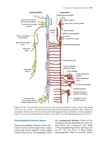
Anatomy of the Nervous System / 211

                              Parasympathetic                Sympathetic
  VetBooks.ir                      Eye     III           Cranial cervical ganglion
                                                               Eye
                      Lacrimal, mandibular,
                      and sublingual glands  VII     Brainstem   Salivary glands
                       Parotid and mucosal  IX                    Sweat and mucosal glands
                            glands         X
                                                  Vertebral      Blood vessels
                                                   nerve
                                                               Middle cervical ganglion
                 Larynx, esophagus,                                Heart
                    and trachea                  Cervical          Bronchi and lungs

                 Recurrent laryngeal                  T1
                      nerve                                     Cervicothoracic ganglion

                        Heart and
                          lungs
                                                 Thoracic


                                                                Sympathetic trunk
                                               Spinal cord      Thoracic (greater)


                         Abdominal                              splanchnic nerve
                          viscera                                   Celiac ganglion
                                                                             Cranial mesenteric
                                                                             ganglion
                                                                             Celiacomesenteric
                                                                             plexus
                                                  Lumbar                     Aorticorenal ganglion


                                                                          Gonadal ganglion
                                                                          Caudal mesenteric
                                                                          ganglion and plexus
                                            S1
                         Pelvic ganglia                                   Hypogastric nerve
                       (part of pelvic plexus                             (to pelvic plexus)
                      to pelvic viscera & colon

               Figure 10-18.  The autonomic nervous system. Left, the parasympathetic outflow (yellow) with cranial
               nerves III, VII, IX, and X and sacral spinal cord segments carrying parasympathetic fibers. Right, the
               sympathetic division (red). Thoracic and cranial lumbar segments make contributions to the sympathetic
               trunk. These nerves are bilateral and are shown on one side only in this drawing for purposes of clarity.


               Parasympathetic Nervous System           the  craniosacral division. Fibers of the
                                                        cranial portion are distributed via four cra-
               The parasympathetic division of the auto-  nial nerves: the oculomotor (III), facial
               nomic nervous system arises from cranial   (VII), glossopharyngeal (IX), and vagus (X)
               nerves  and  sacral  segments  of  the  spinal   nerves. The first three of these supply
               cord; for this reason, it is sometimes called     parasympathetic fibers to smooth muscle