
Page 395 - Anatomy and Physiology of Farm Animals, 8th Edition
P. 395
380 / Anatomy and Physiology of Farm Animals
Esophagus base of the heart, and the esophageal
VetBooks.ir The esophagus is a muscular tube extend hiatus of the diaphragm. Always a seri-
ous condition, choke can be rapidly fatal
ing from the pharynx to the stomach just
caudal to the diaphragm. The end adjacent in ruminants if the obstruction prevents
release of gas (eructation) from the
to the pharynx is kept closed by the m. cri- rumen.
copharyngeus, which passes from its origin
on the cricoid cartilage over the dorsal
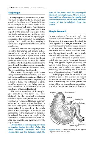
aspect of the proximal esophagus. While Simple Stomach
not in the strictest sense a sphincter mus
cle, the action of the m. cricopharyngeus In nonruminants (horse and pig), the
compresses the opening of the esophagus stomach is just caudal to the left side of the
against the cricoid cartilage, making it diaphragm. It is sometimes described in
function as a sphincter for this end of the these species as a simple stomach. The
esophagus. term “monogastric” is discouraged because
From the pharynx, the esophagus runs it perpetuates the misconception that
dorsal to the trachea and usually inclines ruminants possess more than one stom
somewhat to the left in the neck in the ach; the ruminant actually has a single
midcervical region. It again passes dorsal stomach with multiple compartments.
to the trachea when it enters the thorax The simple stomach is grossly subdi
and continues caudad between the trachea vided into the cardia (entrance), fundus,
and the aorta through the mediastinum to body, and pyloric region (outflow); the
pass through the diaphragm at the esopha- pyloric region features a dense, palpable
geal hiatus. Within the abdominal cavity, sphincter muscle called the pylorus that
the esophagus joins the stomach. controls gastric emptying into more distal
The mucosa of the esophagus is thrown parts of the digestive tract (Fig. 20‐11).
into prominent longitudinal folds that per The esophagus joins the stomach at the
mit considerable cross‐sectional dilation of cardia, a part of the stomach so named
the lumen to accommodate the passage of because of its proximity to the heart. The
a bolus of food. The epithelium is of the walls surrounding the cardia (where the
stratified squamous type, being more or lumen of the esophagus becomes continu
less keratinized in accordance with the ous with that of the stomach) feature a
roughness of the usual feedstuff.
The tunica muscularis of the esopha
gus consists of two layers that cross Fundus Saccus cecus
obliquely in the proximal esophagus,
assume a spiral configuration in the mid‐ Cardia Esophagus
esophageal region, and form an inner cir
cular and an outer longitudinal layer in Pylorus
the more distal parts. The muscle changes
from striated to smooth in the caudal
third of the esophagus in the horse and Body Lesser curvature
Body
just cranial to the diaphragm in the pig; it
Lesser curvature
is striated throughout its length in the
ruminants. Esophageal muscles, both stri
ated and smooth, are innervated by the Greater curvature
vagus nerve.
Duodenum
Obstruction of the esophagus by food
Pyloric region
or foreign bodies is called choke. The
most common locations for such Figure 20-11. External anatomy of the equine
obstruction are the thoracic inlet, the simple stomach. Dorsal view.