
Page 392 - Anatomy and Physiology of Farm Animals, 8th Edition
P. 392
Anatomy of the Digestive System / 377
Tongue
VetBooks.ir The tongue consists of a mass of muscle
covered by mucous membrane. It is divided
into a free apex at the rostral end, a thick 6
body, and a caudal root adjacent to the
pharynx. The entire tongue is mobile 5
through its muscular attachments to the 2
hyoid apparatus and mandible. The muscles
of the tongue (intrinsic muscles) have fib 3
ers oriented in longitudinal, perpendicular, 1
and transverse directions, permitting the
tongue a wide range of movements. This is
particularly evident in the ox, which uses its 4 Root
tongue as a prehensile organ (Fig. 20‐7).
The tongue is covered with thick kerati
nized stratified squamous epithelium. The a
surface is characterized by a large number
of grossly visible projections, the papillae,
which are particularly well developed on the
dorsal surface (Fig. 20‐8). Filiform, fungi b
form, and vallate papillae are found in all
domestic animals, and foliate papillae are
present in the horse, pig, and dog, but not in Body
ruminants. Ruminants additionally have
large conical papillae. The filiform and coni
cal papillae do not bear taste buds (collec
tions of taste cells specialized for gustation;
see Chapter 12), but all other types of papil
lae do. Taste buds may also be found on the
epiglottis, larynx, pharynx, and soft palate. c
The filiform papillae look vaguely
hairlike. In the ox, they consist of a connec
tive tissue core covered by a highly corni
fied epithelial layer. These papillae are d Apex
shorter and softer in the horse than in
other domestic animals, giving the tongue
of the horse its velvety feel. Interspersed
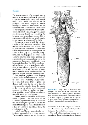
amongst the filiform papillae are fungi- Figure 20-7. Tongue of the ox, dorsal view. The
form papillae, so called because of their pharynx and soft palate are transected and
resemblance to tiny mushrooms. reflected laterad. a, Vallate papillae; b, torus lin
guae with conical papillae; c, fungiform papillae;
Foliate papillae resemble the foliage or d, filiform papillae; 1, oropharynx; 2, tonsillar
leaves of plants. They are found in the crypt; 3, cut surface of soft palate; 4, palatopharyn
horse and pig (and only rarely in cattle) on geal arch; 5, epiglottis; 6, opening into larynx.
the lateral margin adjacent to where the
root of the tongue is connected to the soft
palate by a mucous membrane fold, the the caudal part of the tongue and demar
palatoglossal arch. cate the morphologic division between the
Vallate papillae are large, circular pro body and the root of the tongue.
jections surrounded by a deep groove. The body of the ruminant tongue has a
These papillae are arranged in a V shape on prominent dorsal bulge, the torus lingua,