
Page 389 - Anatomy and Physiology of Farm Animals, 8th Edition
P. 389
374 / Anatomy and Physiology of Farm Animals
membrane of the gum. Some teeth have a covered with enamel, whereas nearly the
entire hypsodont tooth has a layer of
short crown separated from the root by a
VetBooks.ir distinct neck. These teeth, of which rumi enamel, with only a small, short region of
the tooth’s root lacking this layer. The
nant incisors are an example, are described
as brachyodont (from the Greek word enamel of hypsodont teeth is thrown into
brachy, short). In contrast, equine inci prominent folds on the grinding surfaces
sors and cheek teeth have a tall, straight of these teeth, where they form character
crown with no discernible neck. These istic crests (cristae enameli) and cups
teeth are described as hypsodont (Greek (infundibula).
hypsi, high) (Fig. 20‐3). Cementum is a thin, bonelike layer on
Most of the tooth’s substance is made up the surface of the tooth. It covers only the
of a mineralized substance called dentin root of the brachyodont tooth, but it
(sometimes spelled dentine), with a dental extends from the root to cover the crown
cavity at its center. The connective tissues, of the hypsodont tooth.
nerves, and blood vessels of the tooth The front teeth are called incisors, and
reside in this cavity and constitute the in one system of nomenclature they can
dental pulp. It is dentin, incidentally, that be designated by the letter I. The pair of
constitutes the “ivory” of elephant tusks. incisors closest to midline is called I1, or
Superficial to the dentin is a layer of centrals; the next pair I2, or first interme-
enamel, a white layer consisting of inor diates; next I3, or second intermediates;
ganic crystals. Enamel is the hardest sub and the last and most lateral pair of inci
stance in the body. It also is irreplaceable, sors is called I4, or corners. In the nonru
as the cells that generate it (ameloblasts) minants, only one pair of intermediate
are lost following formation of the tooth, incisors is found. Ruminants lack incisors
the only exception amongst farm animals in the upper dental arcade. Instead, the
being the tusks (canine teeth) of swine. mucous membrane in this region is modi
Only the crowns of brachyodont teeth are fied into a dense, keratinized dental pad,
Enamel Infundib ulum
Infundibulum
Crown
Pulp Dentin
cavity Crown
Gingiva
Root
Cementum
Root
Peridontium
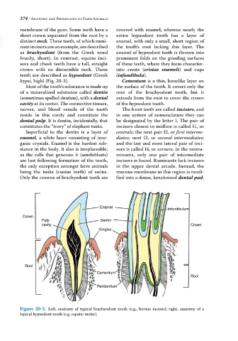
Figure 20-3. Left, anatomy of typical brachyodont tooth (e.g., bovine incisor); right, anatomy of a
typical hypsodont tooth (e.g. equine molar).