
Page 156 - Clinical Small Animal Internal Medicine
P. 156
124 Section 3 Cardiovascular Disease
(holosystolic murmur), or, rarely, throughout systole from Murmurs are traditionally graded on a scale of 1–6.
VetBooks.ir the beginning of S1 to the end of S2 (pansystolic mur- However, evidence exists that a four‐grade scale can be
used for some heart diseases without losing clinically
mur). Diastolic turbulent blood flow is rare in dogs and
cats, and can be relatively difficult to detect. Diastolic
For some cardiac diseases, such as MMVD and aortic/
murmurs most commonly start immediately after the S2. important information (Table 15.1).
Continuous murmurs last throughout systole and dias- pulmonic stenosis, grading of murmurs serves as a rough
tole. The most common heart disease associated with a estimate of disease severity. However, a lack of agreement
continuous murmur is PDA where the murmur com- exists between severity of cardiac disease and murmur
monly peaks in intensity at the time of the S2. grade for other conditions, such as for ventricular septal
Point of maximum intensity (PMI) of a murmur is defects, dilated or restrictive cardiomyopathy (DCM/
commonly the location over the thorax where the mur- RCM), and feline cardiomyopathies. Although the major-
mur is loudest, and can serve as a guide to the origin of ity of dogs with heart disease have an audible murmur, it is
the murmur. The PMI is associated with a particular important to remember that some cardiac diseases (such
valve region (i.e., pulmonic, aortic, mitral, tricuspid). as pericardial diseases, cardiac neoplasia, DCM, atrial sep-
The small size of the feline heart makes it difficult to dis- tal defects [ASD], right‐to‐left shunting PDA, and primary
tinguish the different valve areas on the thorax. myocardial diseases in cats) do not necessarily produce
Accordingly, localization of murmurs in cats is often blood flow turbulence and an audible murmur (Table 15.2).
described as sternal versus parasternal, base versus apex, Presence of a heart murmur does not always signal struc-
and left versus right. The areas of auscultation of the tural cardiac disease as murmurs in cats and dogs can also
heart in dogs are shown in Figure 15.1. The area of mur- arise due to other physiologic processes. Physiologic mur-
mur radiation refers to how widely the murmur can be murs tend to be fairly quiet (grade 1/6–3/6), and may vary in
heard from the PMI, and generally increases with intensity with changes in heart rate. Mild dynamic right ven-
increasing turbulence. tricular outflow tract obstruction is a common cause of mur-
murs in healthy cats in stressful situations such as examination
at a veterinary hospital. Puppies and kittens may have “inno-
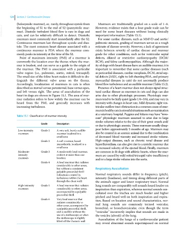
Table 15.1 Classification of murmur intensity
cent” physiologic murmurs assumed to arise due to large
stroke volumes relative to the size of their great vessels and/
Grade Description
or due to physiologic anemia. These murmurs usually disap-
Low‐intensity Grade 1 A very soft, barely audible pear before approximately 5 months of age. Murmurs may
murmurs murmur localized to a also be created in an anemic animal due to the combination
small area of decreased blood viscosity and increased stroke volume.
Grade 2 A soft murmur, heard High‐output diseases, such as chronic renal disease and
immediately, localized to a hyperthyroidism, can also give rise to a systolic murmur due
small area to increased velocity of the ejected blood. Finally, murmurs
Moderate‐ Grade 3 A moderately loud murmur, are common in fit dogs with athletic hearts, where the mur-
intensity evident at more than one murs are caused by mild mitral/tricuspid valve insufficiency
murmurs location and/or a large stroke volume into the aorta.
Grade 4 A loud murmur that radiates
considerably to other areas,
but without a consistent Respiratory Auscultation
palpable precordial thrill
(vibrations created by Normal respiratory sounds differ in frequency (pitch),
turbulence within the heart intensity (loudness), and timing along different parts of
through the chest wall) the animal’s upper and lower respiratory tract. Normal
High‐intensity Grade 5 A loud murmur that radiates lung sounds are comparably soft sounds heard louder on
murmurs considerably to other areas, inspiration than expiration, whereas normal sounds aus-
accompanied by a palpable cultated over the trachea are much louder and higher
precordial thrill pitched and heard well on both inspiration and expira-
Grade 6 Very loud murmur that tion. Based on location and sound characteristics, nor-
radiates considerably to
other areas, accompanied by mal lung sounds are commonly termed vesicular,
a palpable precordial thrill, bronchial, or bronchovesicular, even though the term
and is audible without the “vesicular” incorrectly implies that sounds are made in
use of a stethoscope or when the vesicles (alveoli) of the lung.
the stethoscope is slightly Auscultation of the lungs of a cardiovascular patient
lifted off the thoracic wall
may reveal abnormal sounds superimposed on normal