
Page 160 - Clinical Small Animal Internal Medicine
P. 160
128 Section 3 Cardiovascular Disease
Table 16.1 Main noncardiac‐related factors able to induce cardiac
VetBooks.ir Factors Radiographic change
silhouette changes on thoracic radiographs
Age
Young age (dog) Heart tends to be larger relatively to
the thoracic cavity size than that of
adult dogs
Old age (cat) Heart of cats >7 years old tends to
have a more horizontal orientation on
lateral views, with a prominent aortic
arch on lateral, DV, and VD views
Breed See Table 16.2, text, and Figure 16.2
Patient positioning Chest rotation can increase the
apparent heart base size and that of
the left atrium
Respiration phase Fat
Expiration The heart appears wider than in
inspiration with increased sternal
contact (therefore potentially
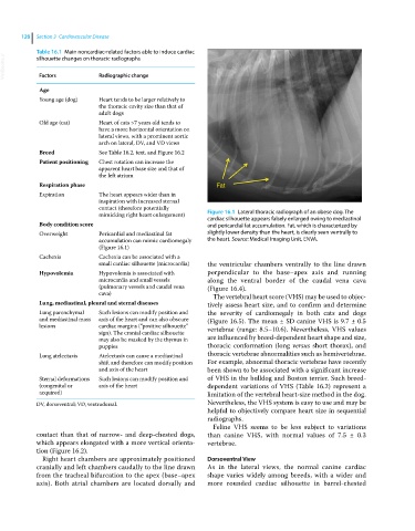
mimicking right heart enlargement) Figure 16.1 Lateral thoracic radiograph of an obese dog. The
cardiac silhouette appears falsely enlarged owing to mediastinal
Body condition score and pericardial fat accumulation. Fat, which is characterized by
Overweight Pericardial and mediastinal fat slightly lower density than the heart, is clearly seen ventrally to
accumulation can mimic cardiomegaly the heart. Source: Medical Imaging Unit, ENVA.
(Figure 16.1)
Cachexia Cachexia can be associated with a
small cardiac silhouette (microcardia) the ventricular chambers ventrally to the line drawn
Hypovolemia Hypovolemia is associated with perpendicular to the base–apex axis and running
microcardia and small vessels along the ventral border of the caudal vena cava
(pulmonary vessels and caudal vena (Figure 16.4).
cava)
The vertebral heart score (VHS) may be used to objec-
Lung, mediastinal, pleural and sternal diseases tively assess heart size, and to confirm and determine
Lung parenchymal Such lesions can modify position and the severity of cardiomegaly in both cats and dogs
and mediastinal mass axis of the heart and can also obscure (Figure 16.5). The mean ± SD canine VHS is 9.7 ± 0.5
lesions cardiac margins (“positive silhouette” vertebrae (range: 8.5–10.6). Nevertheless, VHS values
sign). The cranial cardiac silhouette
may also be masked by the thymus in are influenced by breed‐dependent heart shape and size,
puppies thoracic conformation (long versus short thorax), and
Lung atelectasis Atelectasis can cause a mediastinal thoracic vertebrae abnormalities such as hemivertebrae.
shift and therefore can modify position For example, abnormal thoracic vertebrae have recently
and axis of the heart been shown to be associated with a significant increase
Sternal deformations Such lesions can modify position and of VHS in the bulldog and Boston terrier. Such breed‐
(congenital or axis of the heart dependent variations of VHS (Table 16.2) represent a
acquired) limitation of the vertebral heart‐size method in the dog.
DV, dorsoventral; VD, ventrodorsal. Nevertheless, the VHS system is easy to use and may be
helpful to objectively compare heart size in sequential
radiographs.
Feline VHS seems to be less subject to variations
contact than that of narrow‐ and deep‐chested dogs, than canine VHS, with normal values of 7.5 ± 0.3
which appears elongated with a more vertical orienta- vertebrae.
tion (Figure 16.2).
Right heart chambers are approximately positioned Dorsoventral View
cranially and left chambers caudally to the line drawn As in the lateral views, the normal canine cardiac
from the tracheal bifurcation to the apex (base–apex shape varies widely among breeds, with a wider and
axis). Both atrial chambers are located dorsally and more rounded cardiac silhouette in barrel‐chested