
Page 163 - Clinical Small Animal Internal Medicine
P. 163
16 Imaging in Cardiovascular Disease 131
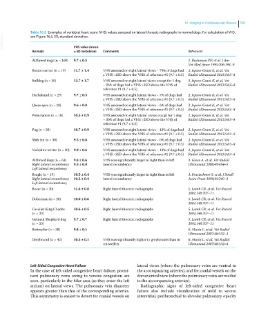
Table 16.2 Examples of vertebral heart score (VHS) values assessed on lateral thoracic radiographs in normal dogs. For calculation of VHS,
VetBooks.ir
see Figure 16.5. SD, standard deviation.
Animals VHS value (mean Comments References
± SD vertebrae)
All breed dogs (n = 100) 9.7 ± 0.5 1. Buchanan JW, et al. J Am
Vet Med Assoc 1995;206:194–9
Boston terrier (n = 19) 11.7 ± 1.4 VHS assessed on right lateral views − 74% of dogs had 2. Jepsen‐Grant K, et al. Vet
a VHS >2SD above the VHS of reference #1 (9.7 ± 0.5) Radiol Ultrasound 2013;54:3–8
Bulldog (n = 30) 12.7 ± 1.7 VHS assessed on right lateral views except for 1 dog 2. Jepsen‐Grant K, et al. Vet
− 93% of dogs had a VHS >2SD above the VHS of Radiol Ultrasound 2013;54:3–8
reference #1 (9.7 ± 0.5)
Dachshund (n = 29) 9.7 ± 0.5 VHS assessed on right lateral views − 7% of dogs had 2. Jepsen‐Grant K, et al. Vet
a VHS >2SD above the VHS of reference #1 (9.7 ± 0.5) Radiol Ultrasound 2013;54:3–8
Lhasa apso (n = 18) 9.6 ± 0.8 VHS assessed on right lateral views − 6% of dogs had 2. Jepsen‐Grant K, et al. Vet
a VHS >2SD above the VHS of reference #1 (9.7 ± 0.5) Radiol Ultrasound 2013;54:3–8
Pomeranian (n = 18) 10.5 ± 0.9 VHS assessed on right lateral views except for 1 dog 2. Jepsen‐Grant K, et al. Vet
− 50% of dogs had a VHS >2SD above the VHS of Radiol Ultrasound 2013;54:3–8
reference #1 (9.7 ± 0.5)
Pug (n = 30) 10.7 ± 0.9 VHS assessed on right lateral views − 43% of dogs had 2. Jepsen‐Grant K, et al. Vet
a VHS >2SD above the VHS of reference #1 (9.7 ± 0.5) Radiol Ultrasound 2013;54:3–8
Shih tzu (n = 30) 9.5 ± 0.6 VHS assessed on right lateral views − 0% of dogs had 2. Jepsen‐Grant K, et al. Vet
a VHS >2SD above the VHS of reference #1 (9.7 ± 0.5) Radiol Ultrasound 2013;54:3–8
Yorkshire terrier (n = 30) 9.9 ± 0.6 VHS assessed on right lateral views − 13% of dogs had 2. Jepsen‐Grant K, et al. Vet
a VHS >2SD above the VHS of reference #1 (9.7 ± 0.5) Radiol Ultrasound 2013;54:3–8
All breed dogs (n = 63) 9.8 ± 0.6 VHS was significantly larger in right than in left 3. Greco A, et al. Vet Radiol
Right lateral recumbency 9.5 ± 0.8 lateral recumbency Ultrasound 2008;49:454–5
Left lateral recumbency
Beagle (n = 19) 10.5 ± 0.4 VHS was significantly larger in right than in left 4. Kraetschmer S, et al. J Small
Right lateral recumbency 10.2 ± 0.4 lateral recumbency Anim Pract 2008;49:240–3
Left lateral recumbency
Boxer (n = 20) 11.6 ± 0.8 Right lateral thoracic radiographs 5. Lamb CR, et al. Vet Record
2001;148:707–11
Doberman (n = 20) 10.0 ± 0.6 Right lateral thoracic radiographs 5. Lamb CR, et al. Vet Record
2001;148:707–11
Cavalier King Charles 10.6 ± 0.5 Right lateral thoracic radiographs 5. Lamb CR, et al. Vet Record
(n = 20) 2001;148:707–11
German Shepherd dog 9.7 ± 0.7 Right lateral thoracic radiographs 5. Lamb CR, et al. Vet Record
(n = 20) 2001;148:707–11
Rottweiler (n = 38) 9.8 ± 0.1 6. Marin L, et al. Vet Radiol
Ultrasound 2007;48:332–4
Greyhound (n = 42) 10.5 ± 0.1 VHS was significantly higher in greyhounds than in 6. Marin L, et al. Vet Radiol
rottweilers Ultrasound 2007;48:332–4
Left‐Sided Congestive Heart Failure lateral views (where the pulmonary veins are ventral to
In the case of left‐sided congestive heart failure, promi- the accompanying arteries), and for caudal vessels on the
nent pulmonary veins owing to venous congestion are dorsoventral view (where the pulmonary veins are medial
seen, particularly in the hilar area (as they enter the left to the accompanying arteries).
atrium) on lateral views. The pulmonary vein diameter Radiographic signs of left‐sided congestive heart
appears greater than that of the corresponding arteries. failure also include visualization of mild to severe
This asymmetry is easiest to detect for cranial vessels on inter stitial, peribronchial to alveolar pulmonary opacity