
Page 168 - Clinical Small Animal Internal Medicine
P. 168
136 Section 3 Cardiovascular Disease
(a) right and left parasternal positions. Transverse views,
VetBooks.ir also called short‐axis views, are obtained with the 2D
ultrasound plane perpendicular to the long axis of the
heart. Conversely, sagittal views, also called long‐axis
views, are obtained from short‐axis views by rotating
the transducer 90° counterclockwise, so that the 2D
ultrasound plane becomes parallel to the long axis of
the heart.
Main Parasternal Short‐Axis Views
The three most commonly used 2D transverse echocar-
diographic views are the right parasternal transven-
tricular short‐axis view (Figure 16.13), the right
parasternal transmitral short‐axis view (Figure 16.14),
(b) and the right parasternal transaortic short‐axis view
(Figure 16.15).
The right parasternal transventricular short‐axis view
(Figure 16.13a) provides transverse visualization of the
two ventricular cavities, and is therefore used to assess
left (Figures 16.13b and 16.13c) and right (Figure 16.13d)
ventricular changes in size and shape. This view is also
commonly used to obtain M‐mode echocardiograms at
the ventricular level.
The right parasternal transmitral short‐axis view
(Figure 16.14) allows visualization of the mitral valve
leaflets within the left ventricular cavity, and is therefore
commonly used to obtain M‐mode echocardiograms at
the mitral valve level.
The right parasternal transaortic short‐axis view
(Figure 16.15a) provides transverse visualization of the
heart base, with the ascending aorta visible as a circle
in the middle of the sector image. This view is com-
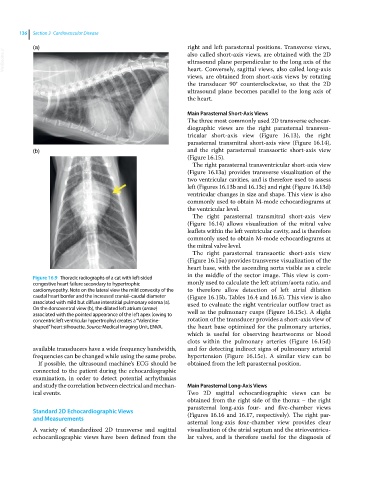
Figure 16.9 Thoracic radiographs of a cat with left‐sided
congestive heart failure secondary to hypertrophic monly used to calculate the left atrium/aorta ratio, and
cardiomyopathy. Note on the lateral view the mild convexity of the to therefore allow detection of left atrial dilation
caudal heart border and the increased cranial–caudal diameter (Figure 16.15b, Tables 16.4 and 16.5). This view is also
associated with mild but diffuse interstitial pulmonary edema (a). used to evaluate the right ventricular outflow tract as
On the dorsoventral view (b), the dilated left atrium (arrow) well as the pulmonary cusps (Figure 16.15c). A slight
associated with the pointed appearance of the left apex (owing to
concentric left ventricular hypertrophy) creates a “Valentine‐ rotation of the transducer provides a short‐axis view of
shaped” heart silhouette. Source: Medical Imaging Unit, ENVA. the heart base optimized for the pulmonary arteries,
which is useful for observing heartworms or blood
clots within the pulmonary arteries (Figure 16.15d)
available transducers have a wide frequency bandwidth, and for detecting indirect signs of pulmonary arterial
frequencies can be changed while using the same probe. hypertension (Figure 16.15e). A similar view can be
If possible, the ultrasound machine’s ECG should be obtained from the left parasternal position.
connected to the patient during the echocardiographic
examination, in order to detect potential arrhythmias
and study the correlation between electrical and mechan- Main Parasternal Long‐Axis Views
ical events. Two 2D sagittal echocardiographic views can be
obtained from the right side of the thorax – the right
parasternal long‐axis four‐ and five‐chamber views
Standard 2D Echocardiographic Views (Figures 16.16 and 16.17, respectively). The right par-
and Measurements
asternal long‐axis four‐chamber view provides clear
A variety of standardized 2D transverse and sagittal visualization of the atrial septum and the atrioventricu-
echocardiographic views have been defined from the lar valves, and is therefore useful for the diagnosis of