
Page 173 - Clinical Small Animal Internal Medicine
P. 173
16 Imaging in Cardiovascular Disease 141
VetBooks.ir (b) (c)
(d)
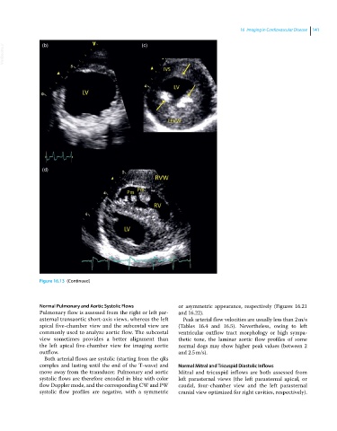
Figure 16.13 (Continued)
Normal Pulmonary and Aortic Systolic Flows or asymmetric appearance, respectively (Figures 16.21
Pulmonary flow is assessed from the right or left par- and 16.22).
asternal transaortic short‐axis views, whereas the left Peak arterial flow velocities are usually less than 2 m/s
apical five‐chamber view and the subcostal view are (Tables 16.4 and 16.5). Nevertheless, owing to left
commonly used to analyze aortic flow. The subcostal ventricular outflow tract morphology or high sympa-
view sometimes provides a better alignment than thetic tone, the laminar aortic flow profiles of some
the left apical five‐chamber view for imaging aortic normal dogs may show higher peak values (between 2
outflow. and 2.5 m/s).
Both arterial flows are systolic (starting from the qRs
complex and lasting until the end of the T‐wave) and Normal Mitral and Tricuspid Diastolic Inflows
move away from the transducer. Pulmonary and aortic Mitral and tricuspid inflows are both assessed from
systolic flows are therefore encoded in blue with color left parasternal views (the left parasternal apical, or
flow Doppler mode, and the corresponding CW and PW caudal, four‐chamber view and the left parasternal
systolic flow profiles are negative, with a symmetric cranial view optimized for right cavities, respectively).