
Page 175 - Clinical Small Animal Internal Medicine
P. 175
16 Imaging in Cardiovascular Disease 143
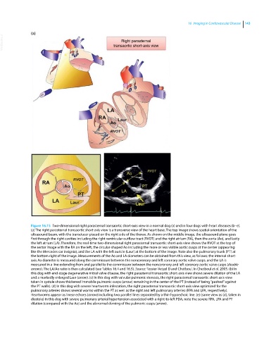
(a)
VetBooks.ir transaortic short-axis view
Right parasternal
Figure 16.15 Two‐dimensional right parasternal transaortic short‐axis view in a normal dog (a) and in four dogs with heart diseases (b–e).
(a) The right parasternal transaortic short‐axis view is a transverse view of the heart base. The top image shows spatial orientation of the
ultrasound beam, with the transducer placed on the right side of the thorax. As shown on the middle image, the ultrasound plane goes
first through the right cavities including the right ventricular outflow tract (RVOT) and the right atrium (RA), then the aorta (Ao), and lastly
the left atrium (LA). Therefore, the real‐time two‐dimensional right parasternal transaortic short‐axis view shows the RVOT at the top of
the sector image with the RA on the left, the circular‐shaped Ao including the more or less visible aortic cusps at the center (appearing
like the Mercedes car insignia), and the LA with the left auricle (Laur) at the bottom of the image. Note also the pulmonary trunk (PT) at
the bottom right of the image. Measurements of the Ao and LA diameters can be obtained from this view, as follows: the internal short‐
axis Ao diameter is measured along the commissure between the noncoronary and left coronary aortic valve cusps, and the LA is
measured in a line extending from and parallel to the commissure between the noncoronary and left coronary aortic valve cusps (double
arrows). The LA/Ao ratio is then calculated (see Tables 16.4 and 16.5). Source: Tessier-Vetzel D and Chetboul. In Chetboul et al. 2005. (b) In
this dog with end‐stage degenerative mitral valve disease, the right parasternal transaortic short‐axis view shows severe dilation of the LA
and a markedly enlarged Laur (arrow). (c) In this dog with valvular pulmonic stenosis, the right parasternal transaortic short‐axis view
taken in systole shows thickened immobile pulmonic cusps (arrow) remaining in the center of the PT (instead of being “pushed” against
the PT walls). (d) In this dog with severe heartworm infestation, the right parasternal transaortic short‐axis view optimized for the
pulmonary arteries shows several worms within the PT as well as the right and left pulmonary arteries (RPA and LPA, respectively).
Heartworms appear as linear echoes (arrow) including two parallel lines separated by a thin hypoechoic line. (e) (same view as (d), taken in
diastole) In this dog with severe pulmonary arterial hypertension associated with a right‐to‐left PDA, note the severe RPA, LPA and PT
dilation (compared with the Ao) and the abnormal doming of the pulmonic cusps (arrow).