
Page 180 - Clinical Small Animal Internal Medicine
P. 180
148 Section 3 Cardiovascular Disease
calculating the maximal ratio of the regurgitant jet area regurgitation. Additionally, the ARJ/LAA ratio may be
VetBooks.ir signal to left atrium area (ARJ/LAA ratio) using color influenced by several factors including systemic arterial
blood pressure, left atrial pressure, and spatial orienta-
flow Doppler mode (Figure 16.30). The major advantage
of this color Doppler mapping method is the rapidity
sible to quantify (rather than “semi‐quantify”) mitral
and ease of data acquisition. Nevertheless, this technique tion of the regurgitant jet. Note that it may also be pos-
presents several limitations: the maximal value for the valve regurgitation (in mL) using another Doppler tech-
ARJ/LAA ratio is 100%, which precludes accurate dis- nique, called the Proximal Isovelocity Surface Area
crimination between dogs with “significant” mitral (PISA) or flow convergence method.
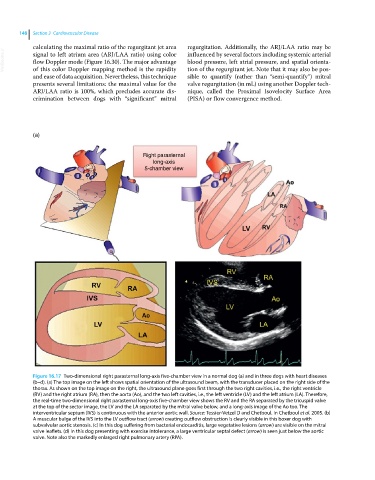
(a)
Right parasternal
long-axis
5-chamber view
Figure 16.17 Two‐dimensional right parasternal long‐axis five‐chamber view in a normal dog (a) and in three dogs with heart diseases
(b–d). (a) The top image on the left shows spatial orientation of the ultrasound beam, with the transducer placed on the right side of the
thorax. As shown on the top image on the right, the ultrasound plane goes first through the two right cavities, i.e., the right ventricle
(RV) and the right atrium (RA), then the aorta (Ao), and the two left cavities, i.e., the left ventricle (LV) and the left atrium (LA). Therefore,
the real‐time two‐dimensional right parasternal long‐axis five‐chamber view shows the RV and the RA separated by the tricuspid valve
at the top of the sector image, the LV and the LA separated by the mitral valve below, and a long‐axis image of the Ao too. The
interventricular septum (IVS) is continuous with the anterior aortic wall. Source: Tessier-Vetzel D and Chetboul. In Chetboul et al. 2005. (b)
A muscular bulge of the IVS into the LV outflow tract (arrow) creating outflow obstruction is clearly visible in this boxer dog with
subvalvular aortic stenosis. (c) In this dog suffering from bacterial endocarditis, large vegetative lesions (arrow) are visible on the mitral
valve leaflets. (d) In this dog presenting with exercise intolerance, a large ventricular septal defect (arrow) is seen just below the aortic
valve. Note also the markedly enlarged right pulmonary artery (RPA).