
Page 182 - Clinical Small Animal Internal Medicine
P. 182
150 Section 3 Cardiovascular Disease
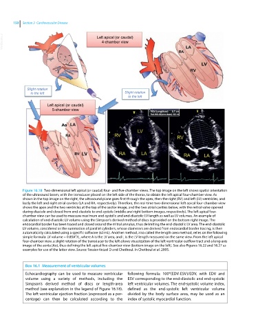
VetBooks.ir Left apical (or caudal)
4-chamber view
Slight rotation
to the left Slight rotation
to the left
Left apical (or caudal)
5-chamber view
Figure 16.18 Two‐dimensional left apical (or caudal) four‐ and five‐chamber views. The top image on the left shows spatial orientation
of the ultrasound beam, with the transducer placed on the left side of the thorax, to obtain the left apical four‐chamber view. As
shown in the top image on the right, the ultrasound plane goes first through the apex, then the right (RV) and left (LV) ventricles, and
lastly the left and right atrial cavities (LA and RA, respectively). Therefore, the real‐time two‐dimensional left apical four‐chamber view
shows the apex and the two ventricles at the top of the sector image, and the two atrial cavities below, with the mitral valve opened
during diastole and closed from end‐diastole to end‐systole (middle and right bottom images, respectively). The left apical four‐
chamber view can be used to measure maximum end‐systolic and end‐diastolic LV length as well as LV volumes. An example of
calculation of end‐diastolic LV volume using the Simpson’s derived method of discs is provided on the bottom right image. The
endocardial border has been traced and closed around the mitral annulus, thus delimiting the end‐diastolic LV area. The end‐diastolic
LV volume, considered as the summation of parallel cylinders, whose diameters are derived from endocardial border tracing, is then
automatically calculated using a specific software (62 mL). Another method, also called the length‐area method, relies on the following
2
simple formula: LV volume = 0.85A /L, where A is the LV area, and L is the LV length measured on the same view. From the left apical
four‐chamber view, a slight rotation of the transducer to the left allows visualization of the left ventricular outflow tract and a long‐axis
image of the aorta (Ao), thus defining the left apical five‐chamber view (bottom image on the left). See also Figures 16.22 and 16.27 as
examples for use of the latter view. Source: Tessier-Vetzel D and Chetboul. In Chetboul et al. 2005.
Box 16.1 Measurement of ventricular volumes
Echocardiography can be used to measure ventricular following formula: 100*(EDV‐ESV)/EDV, with EDV and
volume using a variety of methods, including the ESV corresponding to the end‐diastolic and end‐systolic
Simpson’s derived method of discs or length‐area left ventricular volumes. The end‐systolic volume index,
method (see explanation in the legend of Figure 16.18). defined as the end‐systolic left ventricular volume
The left ventricular ejection fraction (expressed as a per- divided by the body surface area, may be used as an
centage) can then be calculated according to the index of systolic myocardial function.