Page 179 - Clinical Small Animal Internal Medicine
P. 179
16  Imaging in Cardiovascular Disease  147

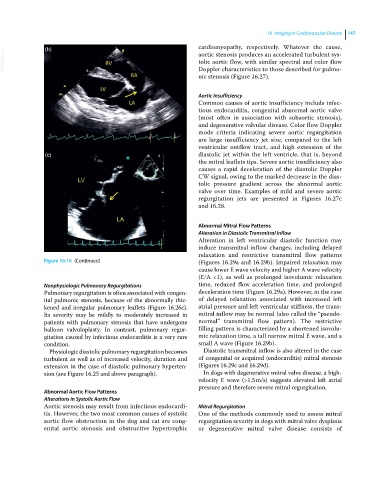
                                                                  cardiomyopathy, respectively. Whatever the cause,
  VetBooks.ir   (b)                                               aortic stenosis produces an accelerated turbulent sys-
                                                                  tolic aortic flow, with similar spectral and color flow
                                                                  Doppler characteristics to those described for pulmo-
                                                                  nic stenosis (Figure 16.27).


                                                                  Aortic Insufficiency
                                                                  Common causes of aortic insufficiency include infec-
                                                                  tious endocarditis, congenital abnormal aortic valve
                                                                  (most often in association with subaortic stenosis),
                                                                  and degenerative valvular disease. Color flow Doppler
                                                                  mode criteria indicating severe aortic regurgitation
                                                                  are large insufficiency jet size, compared to the left
                                                                  ventricular outflow tract, and high extension of the
                (c)                                               diastolic jet within the left ventricle, that is, beyond
                                                                  the mitral leaflets tips. Severe aortic insufficiency also
                                                                  causes a rapid deceleration of the diastolic Doppler
                                                                  CW signal, owing to the marked decrease in the dias-
                                                                  tolic pressure gradient across the abnormal aortic
                                                                  valve over time. Examples of mild and severe aortic
                                                                  regurgitation jets are presented in Figures  16.27c
                                                                  and 16.28.


                                                                  Abnormal Mitral Flow Patterns
                                                                  Alteration in Diastolic Transmitral Inflow
                                                                  Alteration in left ventricular diastolic function may
                                                                  induce transmitral inflow changes, including delayed
                                                                  relaxation and restrictive transmitral flow patterns
               Figure 16.16  (Continued)                          (Figures 16.29a and 16.29b). Impaired relaxation may
                                                                  cause lower E wave velocity and higher A wave velocity
                                                                  (E/A  <1),  as  well  as  prolonged  isovolumic  relaxation
               Nonphysiologic Pulmonary Regurgitations            time, reduced flow acceleration time, and prolonged
               Pulmonary regurgitation is often associated with congen-  deceleration time (Figure 16.29a). However, in the case
               ital pulmonic stenosis, because of the abnormally thic-  of delayed relaxation associated with increased left
               kened and irregular pulmonary leaflets (Figure 16.26c).   atrial pressure and left ventricular stiffness, the trans-
               Its  severity may  be mildly to  moderately increased  in   mitral inflow may be normal (also called the “pseudo-
               patients with pulmonary stenosis that have undergone   normal” transmitral flow pattern). The restrictive
               balloon  valvuloplasty.  In  contrast,  pulmonary  regur-  filling pattern is characterized by a shortened isovolu-
               gitation caused by infectious endocarditis is a very rare   mic relaxation time, a tall narrow mitral E wave, and a
               condition.                                         small A wave (Figure 16.29b).
                 Physiologic diastolic pulmonary regurgitation becomes   Diastolic transmitral inflow is also altered in the case
               turbulent as well as of increased velocity, duration and   of congenital or acquired (endocarditis) mitral stenosis
               extension in the case of diastolic pulmonary hyperten-  (Figures 16.29c and 16.29d).
               sion (see Figure 16.25 and above paragraph).        In dogs with degenerative mitral valve disease, a high‐
                                                                  velocity E wave (>1.5 m/s) suggests elevated left  atrial
                                                                  pressure and therefore severe mitral regurgitation.
               Abnormal Aortic Flow Patterns
               Alterations in Systolic Aortic Flow
               Aortic stenosis may result from infectious endocardi-  Mitral Regurgitation
               tis. However, the two most common causes of systolic   One of the methods commonly used to assess mitral
               aortic flow obstruction in the dog and cat are cong-  regurgitation severity in dogs with mitral valve dysplasia
               enital  aortic  stenosis  and  obstructive  hypertrophic   or degenerative  mitral valve disease consists of

