
Page 181 - Clinical Small Animal Internal Medicine
P. 181
16 Imaging in Cardiovascular Disease 149
(b) (c)
VetBooks.ir
(d)
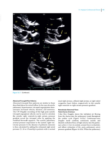
Figure 16.17 (Continued)
Abnormal Tricuspid Flow Patterns sized right atrium, a dilated right atrium, or right‐sided
Abnormal tricuspid flow patterns are similar to those congestive heart failure, respectively) to the systolic
described above for mitral inflow. In the case of systolic right ventricle‐to‐right atrium pressure gradient.
pulmonary hypertension, tricuspid regurgitation dem-
onstrates increased velocity, duration, and extension. Nonvalvular Abnormal Flows
The CW Doppler recording of this high peak systolic Patent Ductus Arteriosus
tricuspid regurgitant velocity can be used to calculate Color flow Doppler shows the turbulent jet flowing
the systolic right ventricle‐to‐right atrium pressure from the ductus into the pulmonary trunk throughout
gradient across the tricuspid valve by applying the the cardiac cycle (Figure 16.31a). Continuous‐wave
modified Bernoulli equation. The systolic pulmonary Doppler mode confirms continuous systolic and
arterial pressure (equivalent to the systolic right ven- diastolic turbulent flows of high velocity into the pulmo-
tricular pressure in the absence of pulmonic stenosis) nary artery (>5 m/s in systole, around 4 m/s in diastole),
is then calculated by adding the estimated right atrial and allows estimation of the aorta–pulmonary artery
pressure (5, 10 or 15 mmHg in patients with a normal pressure gradient (Figure 16.31b). When the pulmonary