
Page 176 - Clinical Small Animal Internal Medicine
P. 176
144 Section 3 Cardiovascular Disease
VetBooks.ir (b) (c)
(d) (e)
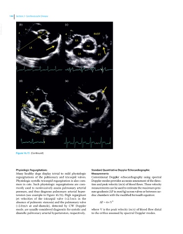
Figure 16.15 (Continued)
Physiologic Regurgitations Standard Quantitative Doppler Echocardiographic
Many healthy dogs display trivial to mild physiologic Measurements
regurgitations of the pulmonary and tricuspid valves. Conventional Doppler echocardiography using spectral
Physiologic systolic tricuspid regurgitation is also com- Doppler modes provides accurate assessment of the direc-
mon in cats. Such physiologic regurgitations are com- tion and peak velocity (m/s) of blood flows. These velocity
monly used to noninvasively assess pulmonary arterial measurements can be used to estimate the maximum pres-
pressure, and thus diagnose pulmonary arterial hyper- sure gradients (∆P in mmHg) across valves or between car-
tension (see example in Figure 16.25). High regurgitant diac chambers with the modified Bernoulli equation:
jet velocities of the tricuspid valve (>2.5 m/s in the
2
absence of pulmonic stenosis) and the pulmonary valve P 4 V
(>2.0 m/s at end-diastole), detected by CW Doppler
mode, are usually considered diagnostic for systolic and where V is the peak velocity (m/s) of blood flow distal
diastolic pulmonary arterial hypertension, respectively. to the orifice assessed by spectral Doppler modes.