
Page 170 - Clinical Small Animal Internal Medicine
P. 170
138 Section 3 Cardiovascular Disease
(a) mation, and has now supplanted invasive techniques
VetBooks.ir such as cardiac catheterization for the diagnosis of most
human and animal heart diseases. Advanced ultrasound
imaging techniques beyond the scope of this chapter are
summarized in Box 16.2.
Basic Principles
Conventional Doppler echocardiography is based on
the Doppler principle, discovered by Christian Doppler
in the 19th century, which states that a moving struc-
ture (e.g., blood cells) reflects the incident ultrasound
(b) beam back to the transducer with a higher or lower
frequency than the original transmitted frequency
(c) depending on whether the structure is, respectively,
moving toward or away from the transducer. This dif-
ference between transmitted and received frequencies,
also called the Doppler shift or frequency shift, is
directly related to the velocity of the reflecting moving
structures, which can thus be deduced from the fre-
Rib 9 quency shift analysis.
The recorded frequency shift (and therefore the calcu-
lated flow velocity) also depends on the cosine of the inter-
cept angle (θ) between the ultrasound beam and blood
flow path. Therefore, whichever Doppler mode is used,
Rib 9
the incident ultrasound beam must be perfectly parallel to
the blood flow path (θ = 0°), otherwise the blood flow
velocity will not be assessed correctly, that is, it will be
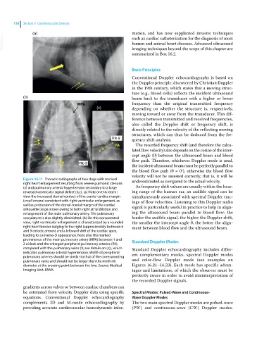
Figure 16.11 Thoracic radiographs of two dogs with marked underestimated as compared to the actual velocity.
right heart enlargement resulting from severe pulmonic stenosis
(a) and pulmonary arterial hypertension secondary to a large As frequency shift values are usually within the hear-
reversed ventricular septal defect (b,c). (a) Note on this lateral ing range of the human ear, an audible signal can be
view the increased sternal contact of the cranial cardiac margin simultaneously associated with spectral Doppler trac-
(small arrows) consistent with right ventricular enlargement, as ings of flow velocities. Listening to this Doppler audio
well as protrusion of the dorsal cranial margin of the cardiac signal is particularly useful in practice to help in align-
silhouette (large arrow) owing to both right atrial dilation and
enlargement of the main pulmonary artery. The pulmonary ing the ultrasound beam parallel to blood flow: the
vasculature is also slightly diminished. (b) On this dorsoventral louder the audible signal, the higher the Doppler shift,
view, right ventricular enlargement is characterized by a rounded the smaller the intercept angle θ, the better the align-
right heart border bulging to the right (approximately between 6 ment between blood flow and the ultrasound beam.
and 9 o’clock; arrows) and a leftward shift of the cardiac apex,
leading to a reverse‐D appearance. Note also the marked
prominence of the main pulmonary artery (MPA) between 1 and Standard Doppler Modes
2 o’clock and the enlarged peripheral pulmonary arteries (PA),
compared with the pulmonary veins (V, see details on (c)), which Standard Doppler echocardiography includes differ-
indicates pulmonary arterial hypertension. Width of peripheral ent complementary modes, spectral Doppler modes
pulmonary arteries should be similar to that of the corresponding
pulmonary veins and should not be larger than the ninth rib and color‐flow Doppler mode (see examples on
diameter at the crossing point between the two. Source: Medical Figures 16.21–16.23). Each mode has specific advan-
Imaging Unit, ENVA. tages and limitations, of which the observer must be
perfectly aware in order to avoid misinterpretation of
the recorded Doppler signals.
gradients across valves or between cardiac chambers can
be estimated from velocity Doppler data using specific Spectral Modes: Pulsed‐Wave and Continuous‐
equations. Conventional Doppler echocardiography Wave Doppler Modes
complements 2D and M‐mode echocardiography by The two main spectral Doppler modes are pulsed‐wave
providing accurate cardiovascular hemodynamic infor- (PW) and continuous‐wave (CW) Doppler modes.