
Page 166 - Clinical Small Animal Internal Medicine
P. 166
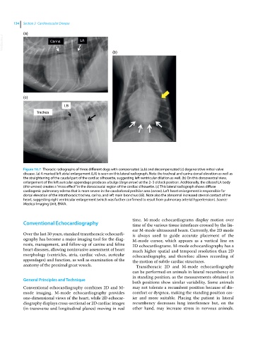
134  Section 3  Cardiovascular Disease

            (a)
  VetBooks.ir           Carina        LA


                                                     (b)


            (c)

                               LB
                  Trachea


            Figure 16.7  Thoracic radiographs of three different dogs with compensated (a,b) and decompensated (c) degenerative mitral valve
            disease. (a) A marked left atrial enlargement (LA) is seen on this lateral radiograph. Note the tracheal and carina dorsal elevation as well as
            the straightening of the caudal part of the cardiac silhouette, suggesting left ventricular dilation as well. (b) On this dorsoventral view,
            enlargement of the left auricular appendage produces a bulge (large arrow) at the 2–3 o’clock position. Additionally, the dilated LA body
            (thin arrows) creates a “mass effect” in the dorsocaudal region of the cardiac silhouette. (c) This lateral radiograph shows diffuse
            cardiogenic pulmonary edema that is more severe in the caudodorsal perihilar area (arrow). Left heart enlargement is responsible for
            dorsal elevation of the intrathoracic trachea, carina, and left main bronchus (LB). Note also the abnormal increased sternal contact of the
            heart, suggesting right ventricular enlargement (which was further confirmed to result from pulmonary arterial hypertension). Source:
            Medical Imaging Unit, ENVA.


                                                              time.  M‐mode  echocardiograms  display  motion  over
              Conventional Echocardiography                   time of the various tissue interfaces crossed by the lin-
                                                              ear M‐mode ultrasound beam. Currently, the 2D mode
            Over the last 30 years, standard transthoracic echocardi-  is always used to guide accurate placement of the
            ography has become a major imaging tool for the diag-  M‐mode cursor, which appears as a vertical line on
            nosis, management, and follow‐up of canine and feline   2D echocardiograms. M‐mode echocardiography has a
            heart diseases, allowing noninvasive assessment of heart   much higher spatial and temporal resolution than 2D
            morphology (ventricles, atria, cardiac valves, auricular   echocardiography, and therefore allows recording of
            appendages) and function, as well as examination of the   the motion of subtle cardiac structures.
            anatomy of the proximal great vessels.              Transthoracic 2D and M‐mode echocardiography
                                                              can be performed on animals in lateral recumbency or
                                                              in standing position, as the measurements obtained in
            General Principles and Technique
                                                              both positions show similar variability. Some animals
            Conventional echocardiography combines 2D and M‐  may not tolerate a recumbent position because of dis-
            mode imaging. M‐mode echocardiography provides    comfort or dyspnea, making the standing position eas-
            one‐dimensional views of the heart, while 2D echocar-  ier and more suitable. Placing the patient in lateral
            diography displays cross‐sectional or 2D cardiac images   recumbency decreases lung interference but, on the
            (in transverse and longitudinal planes) moving in real   other  hand,  may  increase  stress  in  nervous  animals.