
Page 167 - Clinical Small Animal Internal Medicine
P. 167
16 Imaging in Cardiovascular Disease 135
(a)
VetBooks.ir Trachea LB
(c)
1
(b) 2
3
ACDO
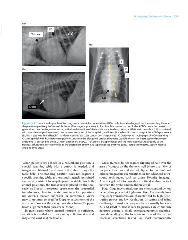
Figure 16.8 Thoracic radiographs of two dogs with patent ductus arteriosus (PDA). (a,b) Lateral radiographs of the same dog (German
shepherd) respectively before and 24 hours after surgery (placement of an Amplatz canine duct occluder, ACDO). Note the marked
generalized heart enlargement on (a), with dorsal elevation of the intrathoracic trachea, carina, and left main bronchus (LB), associated
with vascular congestion (arrows) due to overcirculation of the lung field, and interstitial edema in caudal lungs. After ACDO placement
(b), heart size (width and height) has decreased and vascular congestion disappeared. (c) Dorsoventral radiograph of a Cavalier King
Charles spaniel with PDA before surgical closure. Note the elongated cardiac silhouette (double arrow), the heart base enlargement
(including 1: descending aorta, 2: main pulmonary artery, 3: left auricular appendage), and the increased opacity caudally to the
tracheal bifurcation, corresponding to the dilated left atrium (LA) superimposed over the caudal cardiac silhouette. Source: Medical
Imaging Unit, ENVA.
When patients are echoed in a recumbent position, a Most animals do not require clipping of hair over the
special scanning table with a cutout is needed, and area of contact on the thoracic wall (more than 90% of
images are obtained from beneath the table through the the patients at our unit are not clipped for conventional
table hole. The standing position does not require a echocardiographic examinations or for advanced ultra-
specific scanning table, as the animal is gently restrained sound techniques, such as tissue Doppler imaging).
against an assistant to keep its position stable. For both Acoustic gel helps to provide an optimal air‐free contact
animal positions, the transducer is placed on the tho- between the probe and the thoracic wall.
racic wall in an intercostal space over the precordial High‐frequency transducers are characterized by low
impulse area, close to the sternum, to obtain paraster- penetrating power but high resolution. Conversely, low‐
nal views. However, subcostal (or subxiphoid) views frequency transducers are characterized by high pene-
may sometimes be used for Doppler assessment of the trating power but low resolution. In canine and feline
aortic outflow (as they may provide a better Doppler cardiology, transducer frequencies are usually between
beam alignment than parasternal views). 2.5 and 12 MHz. Transducer frequencies are commonly
In most cases where manual restraint is sufficient, adjusted during a single echocardiographic examina-
sedation is avoided as it can alter systolic function and tion, depending on the location and size of the cardio-
may affect cardiac dimensions. vascular structures tested. As most commercially