
Page 169 - Clinical Small Animal Internal Medicine
P. 169
16 Imaging in Cardiovascular Disease 137
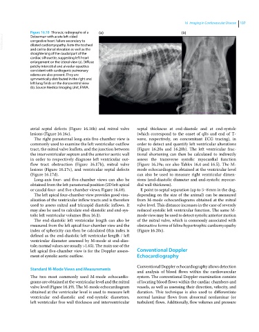
Figure 16.10 Thoracic radiographs of a (a) (b)
VetBooks.ir congestive heart failure secondary to
Doberman with acute left‐sided
dilated cardiomyopathy. Note the tracheal
and carina dorsal elevation as well as the
straightening of the caudal part of the
cardiac silhouette, suggesting left heart
enlargement on the lateral view (a). Diffuse
patchy interstitial and alveolar opacities
consistent with cardiogenic pulmonary
edema are also present. They are
symmetrically distributed in the right and
left lung fields on the dorsoventral view
(b). Source: Medical Imaging Unit, ENVA.
atrial septal defects (Figure 16.16b) and mitral valve septal thickness at end‐diastole and at end‐systole
lesions (Figure 16.16c). (which correspond to the onset of qRs and end of T‐
The right parasternal long‐axis five‐chamber view is wave, respectively, on concomitant ECG tracing), in
commonly used to examine the left ventricular outflow order to detect and quantify left ventricular alterations
tract, the mitral valve leaflets, and the junction between (Figure 16.20a and 16.20b). The left ventricular frac-
the interventricular septum and the anterior aortic wall tional shortening can then be calculated to indirectly
in order to respectively diagnose left ventricular out- assess the transverse systolic myocardial function
flow tract obstruction (Figure 16.17b), mitral valve (Figure 16.19a; see also Tables 16.4 and 16.5). The M‐
lesions (Figure 16.17c), and ventricular septal defects mode echocardiogram obtained at the ventricular level
(Figure 16.17d). can also be used to measure right ventricular dimen-
Long‐axis four‐ and five‐chamber views can also be sions (end‐diastolic diameter and end‐systolic myocar-
obtained from the left parasternal position (2D left apical dial wall thickness).
or caudal four‐ and five‐chamber views; Figure 16.18). E point to septal separation (up to 5–8 mm in the dog,
The left apical four‐chamber view provides good visu- depending on the size of the animal) can be measured
alization of the ventricular inflow tracts and is therefore from M‐mode echocardiograms obtained at the mitral
used to assess mitral and tricuspid diastolic inflows. It valve level. This distance increases in the case of severely
may also be used to calculate end‐diastolic and end‐sys- reduced systolic left ventricular function. The same M‐
tolic left ventricular volumes (Box 16.1). mode view may be used to detect systolic anterior motion
The end‐diastolic left ventricular length can also be of the mitral valve, which is commonly associated with
measured from the left apical four‐chamber view and the obstructive forms of feline hypertrophic cardiomyopathy
index of sphericity can then be calculated (this index is (Figure 16.20c).
defined as the end‐diastolic left ventricular length / left
ventricular diameter assessed by M‐mode at end‐dias-
tole; normal values are usually >1.65). The main use of the
left apical five‐chamber view is for the Doppler assess- Conventional Doppler
ment of systolic aortic outflow. Echocardiography
Conventional Doppler echocardiography allows detection
Standard M‐Mode Views and Measurements
and analysis of blood flows within the cardiovas cular
The two most commonly used M‐mode echocardio- system. The conventional Doppler examination consists
grams are obtained at the ventricular level and the mitral of locating blood flows within the cardiac chambers and
valve level (Figure 16.19). The M‐mode echocardiogram vessels, as well as assessing their direction, velocity, and
obtained at the ventricular level is used to measure left duration. This technique is also used to differentiate
ventricular end‐diastolic and end‐systolic diameters, normal laminar flows from abnormal nonlaminar (or
left ventricular free wall thickness and interventricular turbulent) flows. Additionally, flow volumes and pressure