
Page 164 - Clinical Small Animal Internal Medicine
P. 164
132 Section 3 Cardiovascular Disease
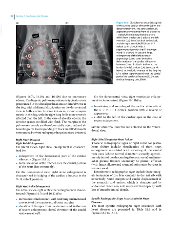
(a) (b) Figure 16.6 Clock face analogy (a) applied
VetBooks.ir dorsoventral view. The aortic arch (AoA)
to the canine cardiac silhouette (b) on the
approximately extends from 11 o’clock to
AoA 1 o’clock, the main pulmonary artery
(MPA) from 1 o’clock to 2 o’clock, the left
MPA ventricle (LV) from 3 o’clock to 6 o’clock,
and the right ventricle (RV) from 6–7
RA LAur o’clock to 11 o’clock (with a
superimposition with the RA between
9 and 11 o’clock). In cats and dogs,
enlargement of the left auricular
appendage (Laur) contributes to a
deformation of the cardiac silhouette
LA
between 2 and 3 o’clock. In the cat, the
body of the left atrium (LA) also extends
from 2 to 3 o’clock, whereas in the dog the
LA is rather superimposed over the caudal
RV LV
part of the cardiac silhouette (b). Source:
Medical Imaging Unit, ENVA.
(Figures 16.7c, 16.10a and 16.10b) due to pulmonary On the dorsoventral view, right ventricular enlarge-
edema. Cardiogenic pulmonary edema is typically more ment is characterized (Figure 16.11b) by:
pronounced in the dorsal perihilar area on lateral views in
the dog, with a bilateral distribution on the dorsoventral ● broadening and rounding of the cardiac silhouette at
view in both species. In some instances, it can be asym- the 6–7 to 9–11 o’clock position with a reverse‐D
metric in the dog, with the right lung fields more severely appearance
affected than the left. In the case of alveolar edema, the ● a shift to the left of the cardiac apex in the case of
alveolar spaces are filled with fluid. The margins of the severe enlargement.
pulmonary vessels are therefore totally obscured and air Similar abnormal patterns are detected on the ventro-
bronchograms (corresponding to black air‐filled bronchi dorsal view.
surrounded by white radiopaque lung tissue) are detected.
Right Heart Diseases Right‐Sided Congestive Heart Failure
Right Atrial Enlargement Thoracic radiographic signs of right‐sided congestive
On lateral views, right atrial enlargement is character- heart failure include visualization of right heart
ized by: enlargement associated with widening of the caudal
vena cava (whose normal diameter is usually approxi-
● enlargement of the dorsocranial part of the cardiac mately that of the descending thoracic aorta) and inter-
silhouette (Figure 16.11a) lobar pleural fissures secondary to pleural effusion
● dorsal elevation of the trachea over the cranial portion (with lung collapse and rounded pulmonary borders in
of the heart (less commonly). severe cases).
On the dorsoventral view, right atrial enlargement is Extrathoracic radiographic signs include hepatomeg-
characterized by bulging of the cardiac silhouette in the aly (extension of the liver caudally to the last rib with
9–11 o’clock position. abnormally round margins and caudal displacement of
the stomach) and ascites, which is characterized by
Right Ventricular Enlargement abdominal distension and increased fluid opacity with
On lateral views, right ventricular enlargement is charac- loss of intraabdominal details.
terized (Figures 16.7c and 16.11a) by:
● increased sternal contact, with widening and increased Specific Radiographic Signs Associated with Heart
convexity of the cranioventral heart margins Diseases
● elevation of the apex from the sternum and, in the case The main specific radiographic signs associated with
of severe enlargement, dorsal elevation of the caudal heart diseases are presented in Table 16.3 and in
vena cava as well. Figures 16.7 to 16.12.